
International
ADVANCED AND APPLIED SCIENCES
EISSN: 2313-3724, Print ISSN: 2313-626X
Frequency: 12
![]()
Volume 12, Issue 11 (November 2025), Pages: 19-28

----------------------------------------------
Original Research Paper
Lung cancer pathological image classification using spatial-channel attention (SCA) mechanism
Author(s):
Affiliation(s):
1Chakrabongse Bhuvanarth International College for Interdisciplinary Studies, Rajamangala University of Technology Tawan-ok, Bangkok, Thailand
2Faculty of Data Science, Guangzhou Huashang College, Guangzhou, China
3Faculty of Data Science, City University of Macau, Macau, China
4Shenzhen Bao’an Chinese Medicine Hospital, Guangzhou University of Chinese Medicine, Shenzhen, China
5Faculty of Business Administration and Information Technology, Rajamangla University of Technology Tawan-ok, Bangkok, Thailand
Full text

* Corresponding Author.
 Corresponding author's ORCID profile: https://orcid.org/0000-0003-0569-0373
Digital Object Identifier (DOI)
https://doi.org/10.21833/ijaas.2025.11.003
Abstract
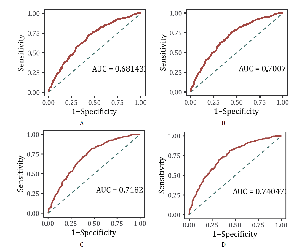
With the increasing use of deep learning in medical imaging, particularly in analyzing lung cancer pathology images, this technology shows great promise for building models for pathological grading and prognosis. This study highlights the growing importance of deep learning in this area, but also notes that accurately classifying lung cancer pathology images remains a difficult task, especially when high precision is needed for grading and prognosis. The aim of this research is to improve the classification of lung cancer pathology images by developing and optimizing a deep learning model. The study focuses on comparing different models, with special attention given to improving the performance of the SCA-ResNet model. The results show that SCA-ResNet performs better than the commonly used ResNet-50 model. It achieves higher scores in several evaluation measures, including precision, recall, specificity, F1 score, and the Kappa coefficient. ROC curve analysis also supports the superior performance of SCA-ResNet, showing better diagnostic accuracy across different cancer grades. These findings suggest that the SCA-ResNet model can offer more accurate and reliable classification of lung cancer pathology images, which may help doctors make better decisions about treatment and prognosis. Its use in clinical practice could lead to improved diagnostic accuracy and better outcomes for patients.
© 2025 The Authors. Published by IASE.
This is an
Keywords
Lung cancer, Deep learning, Pathology images, Image classification, Diagnostic accuracy
Article history
Received 11 February 2025, Received in revised form 23 June 2025, Accepted 9 October 2025
Funding
This research was supported by funding from Guangzhou Huashang College Natural Science Fund of young scholars under grant 2021HSQX53 and Guangzhou Huashang College tutorial system project fund 2022HSDS01. The funding bodies played no role in the design of the study, collection, analysis, or interpretation of data, nor in the writing of the manuscript or the decision to submit it for publication. The authors declare that the content of this manuscript reflects their independent academic work and judgment.
Acknowledgment
No Acknowledgment.
Compliance with ethical standards
Ethical considerations
This study was conducted using the publicly available TCGA-LUAD dataset. All patient data in this resource is fully de-identified and made available with informed consent under controlled access procedures.
Conflict of interest: The author(s) declared no potential conflicts of interest with respect to the research, authorship, and/or publication of this article.
Citation:
Zhong S, Peng Q, Zhang F, Lin W, and Phanniphong K (2025). Lung cancer pathological image classification using spatial-channel attention (SCA) mechanism. International Journal of Advanced and Applied Sciences, 12(11): 19-28
Figures
Fig. 1 Fig. 2 Fig. 3 Fig. 4 Fig. 5
Tables
Table 1 Table 2 Table 3 Table 4 Table 5 Table 6
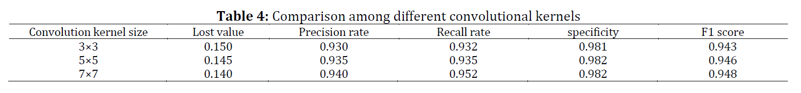
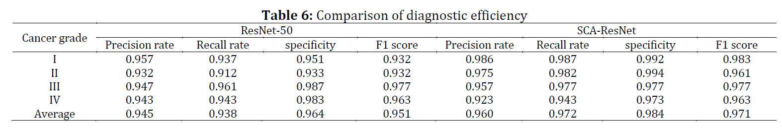
----------------------------------------------
References (23)
- Anand K, Phung TL, Bernicker EH, Cagle PT, Olsen RJ, and Thomas JS (2020). Clinical utility of reflex ordered testing for molecular biomarkers in lung adenocarcinoma. Clinical Lung Cancer, 21(5): 437-442. https://doi.org/10.1016/j.cllc.2020.05.007 [Google Scholar] PMid:32600793
- Baxi V, Edwards R, Montalto M, and Saha S (2022). Digital pathology and artificial intelligence in translational medicine and clinical practice. Modern Pathology, 35(1): 23-32. https://doi.org/10.1038/s41379-021-00919-2 [Google Scholar] PMid:34611303 PMCid:PMC8491759
- Bubendorf L, Lantuejoul S, de Langen AJ, and Thunnissen E (2017). Nonsmall cell lung carcinoma: Diagnostic difficulties in small biopsies and cytological specimens. European Respiratory Review, 26(144): 170007. https://doi.org/10.1183/16000617.0007-2017 [Google Scholar] PMid:28659503 PMCid:PMC9488516
- Civit-Masot J, Bañuls-Beaterio A, Domínguez-Morales M, Rivas-Perez M, Muñoz-Saavedra L, and Corral JMR (2022). Non-small cell lung cancer diagnosis aid with histopathological images using explainable deep learning techniques. Computer Methods and Programs in Biomedicine, 226: 107108. https://doi.org/10.1016/j.cmpb.2022.107108 [Google Scholar] PMid:36113183
- Iqbal MS, Heyat MBB, Parveen S et al. (2024). Progress and trends in neurological disorders research based on deep learning. Computerized Medical Imaging and Graphics, 116: 102400. https://doi.org/10.1016/j.compmedimag.2024.102400 [Google Scholar] PMid:38851079
- Jain DK, Lakshmi KM, Varma KP, Ramachandran M, and Bharati S (2022). Lung cancer detection based on kernel PCA‐convolution neural network feature extraction and classification by fast deep belief neural network in disease management using multimedia data sources. Computational Intelligence and Neuroscience, 2022: 3149406. https://doi.org/10.1155/2022/3149406 [Google Scholar] PMid:35669646 PMCid:PMC9167006
- Kanavati F, Toyokawa G, Momosaki S et al. (2020). Weakly-supervised learning for lung carcinoma classification using deep learning. Scientific Reports, 10: 9297. https://doi.org/10.1038/s41598-020-66333-x [Google Scholar] PMid:32518413 PMCid:PMC7283481
- Kanavati F, Toyokawa G, Momosaki S et al. (2021). A deep learning model for the classification of indeterminate lung carcinoma in biopsy whole slide images. Scientific Reports, 11: 8110. https://doi.org/10.1038/s41598-021-87644-7 [Google Scholar] PMid:33854137 PMCid:PMC8046816
- Kim H, Kwon HJ, Park SY, Park E, and Chung JH (2017). PD-L1 immunohistochemical assays for assessment of therapeutic strategies involving immune checkpoint inhibitors in non-small cell lung cancer: A comparative study. Oncotarget, 8(58): 98524–98532. https://doi.org/10.18632/oncotarget.21567 [Google Scholar] PMid:29228707 PMCid:PMC5716747
- Klupczynska-Gabryszak A, Daskalaki E, Wheelock CE et al. (2024). Metabolomics-based search for lung cancer markers among patients with different smoking status. Scientific Reports, 14: 15444. https://doi.org/10.1038/s41598-024-65835-2 [Google Scholar] PMid:38965272 PMCid:PMC11224321
- Lindeman NI, Cagle PT, Aisner DL et al. (2018). Updated molecular testing guideline for the selection of lung cancer patients for treatment with targeted tyrosine kinase inhibitors: Guideline from the College of American Pathologists, the International Association for the Study of Lung Cancer, and the Association for Molecular Pathology. Archives of Pathology and Laboratory Medicine, 142(3): 321-346. https://doi.org/10.5858/arpa.2017-0388-CP [Google Scholar] PMid:29355391
- Malapelle U, Leprieur EG, Kamga PT, Chiasseu MT, and Rolfo C (2021). Emerging biomarkers for NSCLC: Recent advances in diagnosis and therapy. Frontiers in Oncology, 11: 694578. https://doi.org/10.3389/978-2-88966-915-8 [Google Scholar]
- Moranguinho J, Pereira T, Ramos B, Morgado J, Costa JL, and Oliveira HP (2021). Attention based deep multiple instance learning approach for lung cancer prediction using histopathological images. In the 43rd Annual International Conference of the IEEE Engineering in Medicine and Biology Society, IEEE, Mexico City, Mexico: 2852-2855. https://doi.org/10.1109/EMBC46164.2021.9631000 [Google Scholar] PMid:34891842
- Osmani L, Askin F, Gabrielson E, and Li QK (2018). Current WHO guidelines and the critical role of immunohistochemical markers in the subclassification of non-small cell lung carcinoma (NSCLC): Moving from targeted therapy to immunotherapy. Seminars in Cancer Biology, 52: 103-109. https://doi.org/10.1016/j.semcancer.2017.11.019 [Google Scholar] PMid:29183778 PMCid:PMC5970946
- Ouzzani M, Hammady H, Fedorowicz Z, and Elmagarmid A (2016). Rayyan: A web and mobile app for systematic reviews. Systematic Reviews, 5: 210. https://doi.org/10.1186/s13643-016-0384-4 [Google Scholar] PMid:27919275 PMCid:PMC5139140
- Pan D, Shen J, Al-Huda Z, and Al-Qaness MA (2025). VcaNet: Vision Transformer with fusion channel and spatial attention module for 3D brain tumor segmentation. Computers in Biology and Medicine, 186: 109662. https://doi.org/10.1016/j.compbiomed.2025.109662 [Google Scholar] PMid:39813745
- Rodriguez-Canales J, Parra-Cuentas E, and Wistuba II (2016). Diagnosis and molecular classification of lung cancer. In: Reckamp K (Ed.), Lung cancer: Treatment and research: 25-46. Springer, Cham, Switzerland. https://doi.org/10.1007/978-3-319-40389-2_2 [Google Scholar] PMid:27535388
- Tsuneki M and Kanavati F (2022). Weakly supervised learning for multi-organ adenocarcinoma classification in whole slide images. PLOS ONE, 17(11): e0275378. https://doi.org/10.1371/journal.pone.0275378 [Google Scholar] PMid:36417401 PMCid:PMC9683606
- Udall M, Rizzo M, Kenny J, Doherty J, Dahm S, Robbins P, and Faulkner E (2018). PD-L1 diagnostic tests: A systematic literature review of scoring algorithms and test-validation metrics. Diagnostic Pathology, 13: 12. https://doi.org/10.1186/s13000-018-0689-9 [Google Scholar] PMid:29426340 PMCid:PMC5807740
- Wang S, Yang DM, Rong R et al. (2019). Artificial intelligence in lung cancer pathology image analysis. Cancers, 11(11): 1673. https://doi.org/10.3390/cancers11111673 [Google Scholar] PMid:31661863 PMCid:PMC6895901
- Wen T, Zhang X, Gao Y, Tian H, Fan L, and Yang P (2024). SOX4-BMI1 axis promotes non-small cell lung cancer progression and facilitates angiogenesis by suppressing ZNF24. Cell Death and Disease, 15: 698. https://doi.org/10.1038/s41419-024-07075-w [Google Scholar] PMid:39349443 PMCid:PMC11442842
- Wynes MW, Sholl LM, Dietel M et al. (2014). An international interpretation study using the ALK IHC antibody D5F3 and a sensitive detection kit demonstrates high concordance between ALK IHC and ALK FISH and between evaluators. Journal of Thoracic Oncology, 9(5): 631-638. https://doi.org/10.1097/JTO.0000000000000115 [Google Scholar] PMid:24722153 PMCid:PMC4186652
- Zacharias M, Absenger G, Kashofer K et al. (2021). Reflex testing in non-small cell lung carcinoma using DNA-and RNA-based next-generation sequencing: A single-center experience. Translational Lung Cancer Research, 10(11): 4221–4234. https://doi.org/10.21037/tlcr-21-570 [Google Scholar] PMid:35004252 PMCid:PMC8674594