
International
ADVANCED AND APPLIED SCIENCES
EISSN: 2313-3724, Print ISSN: 2313-626X
Frequency: 12
![]()
Volume 10, Issue 5 (May 2023), Pages: 72-85

----------------------------------------------
Review Paper
Minimal residual disease in acute leukemia based on the insight of molecular genetics monitoring
Author(s):
Najiah M. Alyamani *
Affiliation(s):
Department of Biology, College of Science, University of Jeddah, Jeddah 21493, Saudi Arabia

* Corresponding Author.
 Corresponding author's ORCID profile: https://orcid.org/0000-0002-5457-227X
Digital Object Identifier:
https://doi.org/10.21833/ijaas.2023.05.009
Abstract:
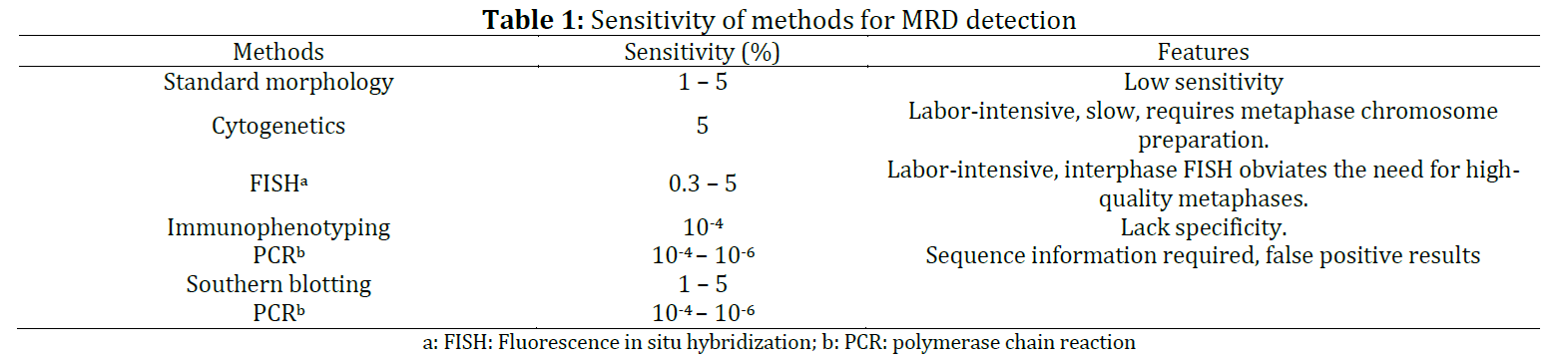
Patients with acute leukemia port 10 malignant cells at presentation. Following chemotherapy or stem cell transplant, patients in complete remission by conventional analyses may still harbor 106/108 malignant cells below the detection limit of standard clinical assessment. Minimal residual disease (MRD) monitoring is one of the most powerful predictors of disease-free and overall survival, particularly for children with acute lymphoblastic leukemia (cALL), the percent annual of cALL increase in the incidence of cALL in Saudi Arabia. Breakpoint fusion regions of chromosomal aberrations can be used as tumor-specific targets for MRD detection by polymerase chain reaction. Levels of MRD, measured at critical time points, significantly correlate with clinical outcomes. Previous works investigated the prognostic significance of leukemia-associated immunophenotypes (LAIPs) as an assessment of the index of MRD in 125 adult B-ALL patients by eight-colour flow cytometry. More advanced molecular and genetics studies are so necessary to identify the mechanisms and cellular structure of the minimal-level disease. Selecting molecular methods for minimal residual disease detection have a much higher sensitivity and precision (100-fold or more) than others. This review highlights the minimal residual disease molecular detection to demonstrate the characterization of the lymphoblastic leukemia gene. Precise MRD monitoring predicts disease relapse after chemotherapy or SCT, provides early intervention, and may result in the rescue of many patients and improvement in the probability of long-term disease-free survival.
© 2023 The Authors. Published by IASE.
This is an
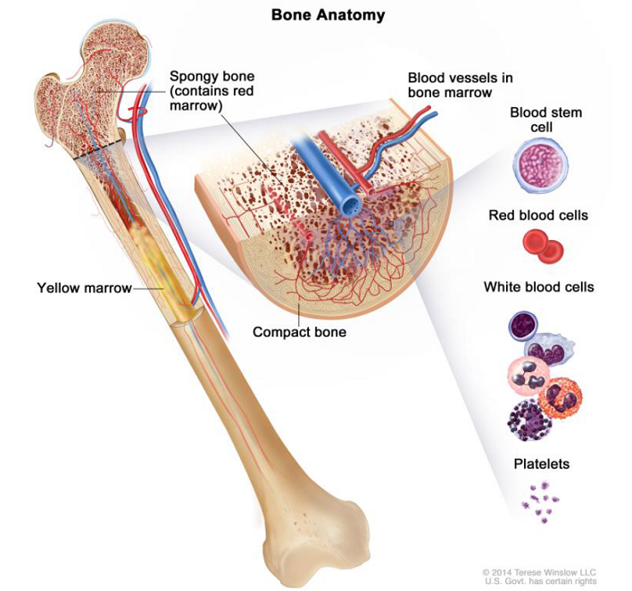
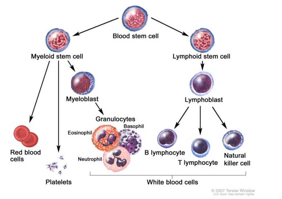
Keywords: Minimal residual disease, Acute lymphoblastic leukemia, Acute myeloid leukemia, Polymerase chain reaction, Bone marrow
Article History: Received 16 November 2022, Received in revised form 10 March 2023, Accepted 14 March 2023
Acknowledgment
No Acknowledgment.
Compliance with ethical standards
Conflict of interest: The author(s) declared no potential conflicts of interest with respect to the research, authorship, and/or publication of this article.
Citation:
Alyamani NM (2023). Minimal residual disease in acute leukemia based on the insight of molecular genetics monitoring. International Journal of Advanced and Applied Sciences, 10(5): 72-85
Figures
Tables
Table 1 Table 2 Table 3 Table 4 Table 5 Table 6 Table 7 Table 8 Table 9 Table 10
----------------------------------------------
References (79)
- Al-Mawali A, Gillis D, and Lewis I (2009). The role of multiparameter flow cytometry for detection of minimal residual disease in acute myeloid leukemia. American Journal of Clinical Pathology, 131(1): 16-26. https://doi.org/10.1309/AJCP5TSD3DZXFLCX [Google Scholar] PMid:19095561
- Anderson K, Lutz C, Van Delft FW, Bateman CM, Guo Y, Colman SM, and Greaves M (2011). Genetic variegation of clonal architecture and propagating cells in leukaemia. Nature, 469(7330): 356-361. https://doi.org/10.1038/nature09650 [Google Scholar] PMid:21160474
- Appelbaum FR (2013). Measurement of minimal residual disease before and after myeloablative hematopoietic cell transplantation for acute leukemia. Best Practice and Research Clinical Haematology, 26(3): 279-284. https://doi.org/10.1016/j.beha.2013.10.008 [Google Scholar] PMid:24309531
- Assumpção JG, Paula FDF, Xavier SG, Murao M, Aguirre Neto JCD, Dutra ÁP, and Viana MB (2013). Gene rearrangement study for minimal residual disease monitoring in children with acute lymphocytic leukemia. Revista Brasileira de Hematologia e Hemoterapia, 35(5): 337-342. https://doi.org/10.5581/1516-8484.20130115 [Google Scholar] PMid:24255617 PMCid:PMC3832314
- Barragan E, Bolufer P, Moreno I, Martin G, Nomdedeu J, Brunet S, and Sanz MA (2001). Quantitative detection of AML1-ETO rearrangement by real-time RT-PCR using fluorescently labeled probes. Leukemia and Lymphoma, 42(4): 747-756. https://doi.org/10.3109/10428190109099337 [Google Scholar] PMid:11697505
- Beishuizen A, Verhoeven MA, Mol EJ, Breit TM, Wolvers-Tettero ILM, and van Dongen JJM (1993). Detection of immunoglobulin heavy-chain gene rearrangements by Southern blot analysis: Recommendations for optimal results. Leukemia, 7: 2045-2053. [Google Scholar] PMid:7902888
- Board PPTE (2021). Childhood acute lymphoblastic leukemia treatment (PDQ®). In PDQ Cancer Information Summaries, National Cancer Institute (US), Rockville, USA. [Google Scholar]
- Boeckx N, Willemse MJ, Szczepanski T, van der Velden VH, Langerak AW, Vandekerckhove P, and van Dongen JJ (2002). Fusion gene transcripts and Ig/TCR gene rearrangements are complementary but infrequent targets for PCR-based detection of minimal residual disease in acute myeloid leukemia. Leukemia, 16(3): 368-375. https://doi.org/10.1038/sj.leu.2402387 [Google Scholar] PMid:11896540
- Boldeanu F, Ordodi VL, Gruia A, Cristea M, Gai E, and Serban M (2011). Minimal residual disease-generalities and perspectives. Timisoara Medical Journal, 61: 3-4. [Google Scholar]
- Buonamici S, Ottaviani E, Testoni N, Montefusco V, Visani G, Bonifazi F, and Martinelli G (2002). Real-time quantitation of minimal residual disease in inv (16)-positive acute myeloid leukemia may indicate risk for clinical relapse and may identify patients in a curable state. Blood, The Journal of the American Society of Hematology, 99(2): 443-449. https://doi.org/10.1182/blood.V99.2.443 [Google Scholar] PMid:11781223
- Campana D (2004). Minimal residual disease studies in acute leukemia. Pathology Patterns Reviews, 122(suppl_1): S47-S57. https://doi.org/10.1309/YJP8CEQ1E7RYP3MF [Google Scholar] PMid:15690642
- Campana D (2010). Minimal residual disease in acute lymphoblastic leukemia. Hematology 2010, the American Society of Hematology Education Program, 2010(1): 7-12. https://doi.org/10.1182/asheducation-2010.1.7 [Google Scholar] PMid:21239764
- Campana D and Pui CH (2008). Childhood leukemia. In: Abeloff MD, Armitage JO, Niederhuber JE, Kastan MB, and McKenna WG (Eds.), Abeloff’s clinical oncology. 4th Edition, Elsevier; Philadelphia, USA. [Google Scholar]
- Chen JS, Coustan-Smith E, Suzuki T, Neale GA, Mihara K, Pui CH, and Campana D (2001). Identification of novel markers for monitoring minimal residual disease in acute lymphoblastic leukemia. Blood: The Journal of the American Society of Hematology, 97(7): 2115-2120. https://doi.org/10.1182/blood.V97.7.2115 [Google Scholar] PMid:11264179
- Cheng SH, Lau KM, Li CK, Chan NP, Ip RK, Cheng CK, and Ng MH (2013). Minimal residual disease-based risk stratification in Chinese childhood acute lymphoblastic leukemia by flow cytometry and plasma DNA quantitative polymerase chain reaction. PLOS ONE, 8(7): e69467. https://doi.org/10.1371/journal.pone.0069467 [Google Scholar] PMid:23936021 PMCid:PMC3723913
- Cirmena G, Ferrando L, Ravera F, Garuti A, Dameri M, Gallo M, and Zoppoli G (2022). Plasma cell-free DNA integrity assessed by automated electrophoresis predicts the achievement of pathologic complete response to Neoadjuvant chemotherapy in patients with breast cancer. JCO Precision Oncology, 6: e2100198. https://doi.org/10.1200/PO.21.00198 [Google Scholar] PMid:35201850 PMCid:PMC8974578
- Coustan-Smith E, Behm FG, Sanchez J, Boyett JM, Hancock ML, Raimondi SC, and Campana D (1998). Immunological detection of minimal residual disease in children with acute lymphoblastic leukaemia. The Lancet, 351(9102): 550-554. https://doi.org/10.1016/S0140-6736(97)10295-1 [Google Scholar] PMid:9492773
- Coustan-Smith E, Song G, Clark C, Key L, Liu P, Mehrpooya M, and Campana D (2011). New markers for minimal residual disease detection in acute lymphoblastic leukemia. Blood: The Journal of the American Society of Hematology, 117(23): 6267-6276. https://doi.org/10.1182/blood-2010-12-324004 [Google Scholar] PMid:21487112 PMCid:PMC3122946
- Czyz A and Nagler A (2019). The role of measurable residual disease (MRD) in hematopoietic stem cell transplantation for hematological malignancies focusing on acute leukemia. International Journal of Molecular Sciences, 20(21): 5362. https://doi.org/10.3390/ijms20215362 [Google Scholar] PMid:31661875 PMCid:PMC6862140
- De Bie J, Demeyer S, Alberti-Servera L, Geerdens E, Segers H, Broux M, and Cools J (2018). Single-cell sequencing reveals the origin and the order of mutation acquisition in T-cell acute lymphoblastic leukemia. Leukemia, 32(6): 1358-1369. https://doi.org/10.1038/s41375-018-0127-8 [Google Scholar] PMid:29740158 PMCid:PMC5990522
- Della Starza I, Chiaretti S, De Propris MS, Elia L, Cavalli M, De Novi LA, and Foà R (2019). Minimal residual disease in acute lymphoblastic leukemia: Technical and clinical advances. Frontiers in Oncology, 9: 726. https://doi.org/10.3389/fonc.2019.00726 [Google Scholar] PMid:31448230 PMCid:PMC6692455
- Dobson SM, García-Prat L, Vanner RJ, Wintersinger J, Waanders E, Gu Z, and Dick JE (2020). Relapse-fated latent diagnosis subclones in acute B lineage leukemia are drug tolerant and possess distinct metabolic programscharacterization of relapse-fated clones in diagnosis B-ALL. Cancer Discovery, 10(4): 568-587. https://doi.org/10.1158/2159-8290.CD-19-1059 [Google Scholar] PMid:32086311 PMCid:PMC7122013
- Feller N, Van Der Velden VHJ, Brooimans RA, Boeckx N, Preijers F, Kelder A, and Schuurhuis GJ (2013). Defining consensus leukemia-associated immunophenotypes for detection of minimal residual disease in acute myeloid leukemia in a multicenter setting. Blood Cancer Journal, 3: e129. https://doi.org/10.1038/bcj.2013.27 [Google Scholar] PMid:23912609 PMCid:PMC3763381
- Foroni L, Gameiro PM, and Hoffbrand V (2005). Minimal residual disease in acute leukemia. In: Hoffbrand AV, Catovsky D, and Tuddenham EGD (Eds.), Postgraduate haematology. 5th Edition, Blackwell Publishing, Hoboken, USA. [Google Scholar]
- Gabert J, Beillard E, Van der Velden VHJ, Bi W, Grimwade D, Pallisgaard N, and Van Dongen JJM (2003). Standardization and quality control studies of ‘real-time’ quantitative reverse transcriptase polymerase chain reaction of fusion gene transcripts for residual disease detection in leukemia–A Europe against cancer program. Leukemia, 17(12): 2318-2357. https://doi.org/10.1038/sj.leu.2403135 [Google Scholar] PMid:14562125
- Gao YJ, He YJ, Yang ZL, Shao HY, Zuo Y, Bai Y, and Zhang L (2010). Increased integrity of circulating cell-free DNA in plasma of patients with acute leukemia. Clinical Chemistry and Laboratory Medicine, 48(11): 1651-1656. https://doi.org/10.1515/CCLM.2010.311 [Google Scholar] PMid:20831457
- Gotham D, McKenna L, Deborggraeve S, Madoori S, and Branigan D (2021). Public investments in the development of GeneXpert molecular diagnostic technology. PLOS ONE, 16(8): e0256883. https://doi.org/10.1371/journal.pone.0256883 [Google Scholar] PMid:34464413 PMCid:PMC8407584
- Guerrasio A, Pilatrino C, De Micheli D, Cilloni D, Serra A, Gottardi E, and Saglio G (2002). Assessment of minimal residual disease (MRD) in CBFbeta/MYH11-positive acute myeloid leukemias by qualitative and quantitative RT-PCR amplification of fusion transcripts. Leukemia, 16(6): 1176-1181. https://doi.org/10.1038/sj.leu.2402478 [Google Scholar] PMid:12040450
- Hackl H, Astanina K, and Wieser R (2017). Molecular and genetic alterations associated with therapy resistance and relapse of acute myeloid leukemia. Journal of Hematology and Oncology, 10: 51. https://doi.org/10.1186/s13045-017-0416-0 [Google Scholar] PMid:28219393 PMCid:PMC5322789
- Hoelzer D and Gökbuget N (2000a). Adult acute lymphoblastic leukemia. In: Hoffbrand AV, Catovsky D, and Tuddenham EGD (Eds.), Postgraduate haematology. 5th Edition, Blackwell Publishing, Hoboken, USA. [Google Scholar]
- Hoelzer D and Gökbuget N (2000b). Recent approaches in acute lymphoblastic leukemia in adults. Critical Reviews in Oncology/Hematology, 36(1): 49-58. https://doi.org/10.1016/S1040-8428(00)00097-4 [Google Scholar] PMid:10996522
- Hoyos M, Nomdedeu JF, Esteve J, Duarte R, Ribera JM, Llorente A, and Sierra J (2013). Core binding factor acute myeloid leukemia: The impact of age, leukocyte count, molecular findings, and minimal residual disease. European Journal of Haematology, 91(3): 209-218. https://doi.org/10.1111/ejh.12130 [Google Scholar] PMid:23646898
- Iacobucci I and Mullighan CG (2017). Genetic basis of acute lymphoblastic leukemia. Journal of Clinical Oncology, 35(9): 975-983. https://doi.org/10.1200/JCO.2016.70.7836 [Google Scholar] PMid:28297628 PMCid:PMC5455679
- Inaba H and Mullighan CG (2020). Pediatric acute lymphoblastic leukemia. Haematologica, 105(11): 2524-2539. https://doi.org/10.3324/haematol.2020.247031 [Google Scholar] PMid:33054110 PMCid:PMC7604619
- Iñigo SDLI, Casares MTG, Jorge CEL, Brito JL, and Cabrera PM (2011). New molecular markers in acute myeloid leukemia. In: Koschmieder S and Krug U (Eds.), Myeloid leukemia-basic mechanisms of leukemogenesis: 312-338. IntechOpen, London, UK. [Google Scholar]
- Jourdan E, Boissel N, Chevret S, Delabesse E, Renneville A, Cornillet P, and Dombret H (2013). Prospective evaluation of gene mutations and minimal residual disease in patients with core binding factor acute myeloid leukemia. Blood: The Journal of the American Society of Hematology, 121(12): 2213-2223. https://doi.org/10.1182/blood-2012-10-462879 [Google Scholar] PMid:23321257
- Juárez-Avendaño G, Méndez-Ramírez N, Luna-Silva NC, Gómez-Almaguer D, Pelayo R, and Balandrán JC (2021). Molecular and cellular markers for measurable residual disease in acute lymphoblastic leukemia. Boletín médico del Hospital Infantil de México, 78(3): 159-170. https://doi.org/10.24875/BMHIM.20000155 [Google Scholar] PMid:34167145
- Kim IS (2020). Minimal residual disease in acute lymphoblastic leukemia: Technical aspects and implications for clinical interpretation. Blood Research, 55(S1): S19-S26. https://doi.org/10.5045/br.2020.S004 [Google Scholar] PMid:32719172 PMCid:PMC7386891
- Kruse A, Abdel-Azim N, Kim HN, Ruan Y, Phan V, Ogana H, and Kim YM (2020). Minimal residual disease detection in acute lymphoblastic leukemia. International Journal of Molecular Sciences, 21(3): 1054. https://doi.org/10.3390/ijms21031054 [Google Scholar] PMid:32033444 PMCid:PMC7037356
- Li B, Brady SW, Ma X, Shen S, Zhang Y, Li Y, and Zhang J et al. (2020). Therapy-induced mutations drive the genomic landscape of relapsed acute lymphoblastic leukemia. Blood, 135(1): 41-55. https://doi.org/10.1182/blood.2019002220 [Google Scholar] PMid:31697823 PMCid:PMC6940198
- Li B, Li H, Bai Y, Kirschner-Schwabe R, Yang JJ, Chen Y, and Zhou BBS (2015). Negative feedback–defective PRPS1 mutants drive thiopurine resistance in relapsed childhood ALL. Nature Medicine, 21(6): 563-571. https://doi.org/10.1038/nm.3840 [Google Scholar] PMid:25962120 PMCid:PMC4670083
- Liang D and Pui C (2000). Childhood acute lymphoblastic leukemia. In: Hoffbrand AV, Catovsky D, and Tuddenham EGD (Eds.), Postgraduate haematology. 5th Edition, Blackwell Publishing, Hoboken, USA. [Google Scholar]
- Ma X, Edmonson M, Yergeau D, Muzny DM, Hampton OA, Rusch M, and Zhang J (2015). Rise and fall of subclones from diagnosis to relapse in pediatric B-acute lymphoblastic leukaemia. Nature Communications, 6: 6604. https://doi.org/10.1038/ncomms7604 [Google Scholar] PMid:25790293 PMCid:PMC4377644
- Meyer JA, Wang J, Hogan LE, Yang JJ, Dandekar S, Patel JP, and Carroll WL (2013). Relapse-specific mutations in NT5C2 in childhood acute lymphoblastic leukemia. Nature Genetics, 45(3): 290-294. https://doi.org/10.1038/ng.2558 [Google Scholar] PMid:23377183 PMCid:PMC3681285
- Möricke A, Zimmermann M, Reiter A, Henze G, Schrauder A, Gadner H, and Schrappe M (2010). Long-term results of five consecutive trials in childhood acute lymphoblastic leukemia performed by the ALL-BFM study group from 1981 to 2000. Leukemia, 24(2): 265-284. https://doi.org/10.1038/leu.2009.257 [Google Scholar] PMid:20010625
- Mukda E, Pintaraks K, Komvilaisak P, and Wiangnon S (2011). Cytochemistry and multi-color flow cytometric immunophenotype for diagnosis of childhood acute leukemia. Journal of Hematology and Transfusion Medicine, 21(1): 23-32. [Google Scholar]
- Paietta E (2012). Minimal residual disease in acute myeloid leukemia: Coming of age. Hematology 2010, the American Society of Hematology Education Program, 2012(1): 35-42. https://doi.org/10.1182/asheducation.V2012.1.35.3797926 [Google Scholar] PMid:23233558
- Peters JM and Ansari MQ (2011). Multiparameter flow cytometry in the diagnosis and management of acute leukemia. Archives of Pathology and Laboratory Medicine, 135(1): 44-54. https://doi.org/10.5858/2010-0387-RAR.1 [Google Scholar] PMid:21204710
- Pongers-Willemse MJ, Seriu T, Stolz F, d’Aniello E, Gameiro P, Pisa P, and Van Dongen JJM (1999). Primers and protocols for standardized detection of minimal residual disease in acute lymphoblastic leukemia using immunoglobulin and T cell receptor gene rearrangements and TAL1 deletions as PCR targets Report of the BIOMED-1 CONCERTED ACTION: Investigation of minimal residual disease in acute leukemia. Leukemia, 13(1): 110-118. https://doi.org/10.1038/sj.leu.2401245 [Google Scholar] PMid:10049045
- Pott C, Brüggemann M, Ritgen M, van der Velden VH, van Dongen JJ, and Kneba M (2019). MRD detection in B-cell non-Hodgkin lymphomas using Ig gene rearrangements and chromosomal translocations as targets for real-time quantitative PCR. Lymphoma: Methods and Protocols, 1956: 199-228. https://doi.org/10.1007/978-1-4939-9151-8_9 [Google Scholar] PMid:30779036
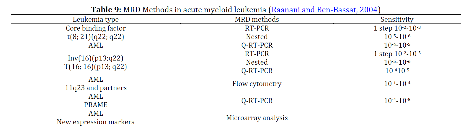
- Raanani P and Ben-Bassat I (2004). Detection of minimal residual disease in acute myelogenous leukemia. Acta Haematologica, 112(1-2): 40-54. https://doi.org/10.1159/000077559 [Google Scholar] PMid:15179004
- Reiter A, Schrappe M, Ludwig WD, Hiddemann W, Sauter S, Henze G, and Riehm H (1994). Chemotherapy in 998 unselected childhood acute lymphoblastic leukemia patients: Results and conclusions of the multicenter trial ALL-BFM 86. Blood, 84(9): 3122-3133. https://doi.org/10.1182/blood.V84.9.3122.bloodjournal8493122 [Google Scholar] PMid:7949185
- Roloff GW, Lai C, Hourigan CS, and Dillon LW (2017). Technical advances in the measurement of residual disease in acute myeloid leukemia. Journal of Clinical Medicine, 6(9): 87. https://doi.org/10.3390/jcm6090087 [Google Scholar] PMid:28925935 PMCid:PMC5615280
- Rosenberg AS, Brunson A, Paulus JK, Tuscano J, Wun T, Keegan THM, and Jonas BA (2017). Secondary acute lymphoblastic leukemia is a distinct clinical entity with prognostic significance. Blood Cancer Journal, 7: e605. https://doi.org/10.1038/bcj.2017.81 [Google Scholar] PMid:28885611 PMCid:PMC5709750
- Samra MA, Mahmoud HK, Abdelhamid TM, El Sharkawy NM, Elnahass YH, Elgammal M, and Kamel AM (2013). The prognostic significance of minimal residual disease in adult Egyptian patients with precursor acute lymphoblastic leukemia. Journal of the Egyptian National Cancer Institute, 25(3): 135-142. https://doi.org/10.1016/j.jnci.2013.05.004 [Google Scholar] PMid:23932750
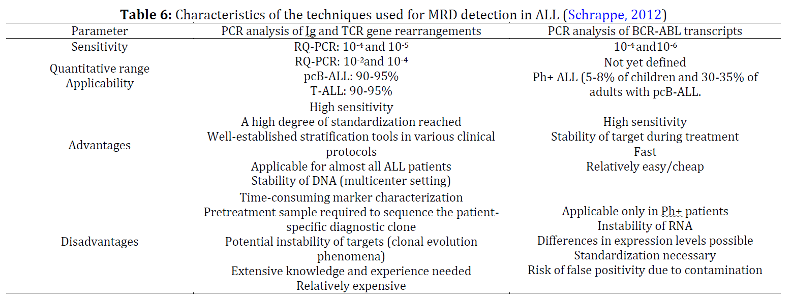
- Schrappe M (2012). Minimal residual disease: Optimal methods, timing, and clinical relevance for an individual patient. Hematology 2010, the American Society of Hematology Education Program, 2012(1): 137-142. https://doi.org/10.1182/asheducation.V2012.1.137.3798216 [Google Scholar] PMid:23233572
- Schuurhuis GJ, Cloos J, and Ossenkoppele GJ (2013). How to assess minimal residual disease in pediatric and adult acute myeloid leukemia? Translational Pediatrics, 2(2): 80-83. [Google Scholar]
- Shayegi N, Kramer M, Bornhäuser M, Schaich M, Schetelig J, Platzbecker U, and Thiede C (2013). The level of residual disease based on mutant NPM1 is an independent prognostic factor for relapse and survival in AML. Blood: The Journal of the American Society of Hematology, 122(1): 83-92. https://doi.org/10.1182/blood-2012-10-461749 [Google Scholar] PMid:23656730
- Shook D, Coustan-Smith E, Ribeiro RC, Rubnitz JE, and Campana D (2009). Minimal residual disease quantitation in acute myeloid leukemia. Clinical Lymphoma and Myeloma, 9(Supplement 3): S281-S285. https://doi.org/10.3816/CLM.2009.s.024 [Google Scholar] PMid:19778853 PMCid:PMC2785493
- Short NJ and Jabbour E (2017). Minimal residual disease in acute lymphoblastic leukemia: How to recognize and treat it. Current Oncology Reports, 19: 6. https://doi.org/10.1007/s11912-017-0565-x [Google Scholar] PMid:28205134
- Sievers EL, Lange BJ, Alonzo TA, Gerbing RB, Bernstein ID, Smith FO, and Loken MR (2003). Immunophenotypic evidence of leukemia after induction therapy predicts relapse: Results from a prospective Children's Cancer Group study of 252 patients with acute myeloid leukemia. Blood: The Journal of the American Society of Hematology, 101(9): 3398-3406. https://doi.org/10.1182/blood-2002-10-3064 [Google Scholar] PMid:12506020
- Sommer U, Eck S, Marszalek L, Stewart JJ, Bradford J, McCloskey TW, and Litwin V (2021). High‐sensitivity flow cytometric assays: Considerations for design control and analytical validation for identification of rare events. Cytometry Part B: Clinical Cytometry, 100(1): 42-51. https://doi.org/10.1002/cyto.b.21949 [Google Scholar] PMid:32940947
- Sun NN, Gan SL, Sun H, Zhang QT, Liu YF, and Xie XS (2013). Dynamically monitoring minimal residual disease in acute leukemia after complete remission by multiparameter flow cytometry and its relation with prognosis. Zhongguo Shi Yan Xue Ye Xue Za Zhi, 21(2): 339-342. https://doi.org/10.7534/j.issn.1009-2137.2013.02.016 [Google Scholar] PMid:23628028
- Szczepański T (2007). Why and how to quantify minimal residual disease in acute lymphoblastic leukemia? Leukemia, 21(4): 622-626. https://doi.org/10.1038/sj.leu.2404603 [Google Scholar] PMid:17301806
- Szczepański T, Willemse MJ, Kamps WA, van Wering ER, Langerak AW, and van Dongen JJ (2001a). Molecular discrimination between relapsed and secondary acute lymphoblastic leukemia: Proposal for an easy strategy. Medical and Pediatric Oncology: The Official Journal of SIOP-International Society of Pediatric Oncology (Societé Internationale d'Oncologie Pédiatrique, 36(3): 352-358. https://doi.org/10.1002/mpo.1085 [Google Scholar] PMid:11241436
- Szczepański T, Willemse MJ, Van Wering ER, Van Weerden JF, Kamps WA, and Van Dongen JJM (2001b). Precursor-B-ALL with DH–JH gene rearrangements have an immature immunogenotype with a high frequency of oligoclonality and hyperdiploidy of chromosome 14. Leukemia, 15(9): 1415-1423. https://doi.org/10.1038/sj.leu.2402206 [Google Scholar] PMid:11516102
- Terwijn M, van Putten WL, Kelder A, van der Velden VH, Brooimans RA, Pabst T, and Schuurhuis GJ (2013). High prognostic impact of flow cytometric minimal residual disease detection in acute myeloid leukemia: Data from the HOVON/SAKK AML 42A study. Journal of Clinical Oncology, 31(31): 3889-3897. https://doi.org/10.1200/JCO.2012.45.9628 [Google Scholar] PMid:24062400
- van der Burg M, Barendregt BH, Szczepanski T, Van Wering ER, Langerak AW, and Van Dongen JJM (2002). Immunoglobulin light chain gene rearrangements display hierarchy in absence of selection for functionality in precursor-B-ALL. Leukemia, 16(8): 1448-1453. https://doi.org/10.1038/sj.leu.2402548 [Google Scholar] PMid:12145684
- van der Reijden BA, Simons A, Luiten E, Van Der Poel SC, Hogenbirk PE, Tönnissen E, and Jansen JH (2002). Minimal residual disease quantification in patients with acute myeloid leukaemia and inv (16)/CBFB‐MYH11 gene fusion. British Journal of Haematology, 118(2): 411-418. https://doi.org/10.1046/j.1365-2141.2002.03738.x [Google Scholar] PMid:12139724
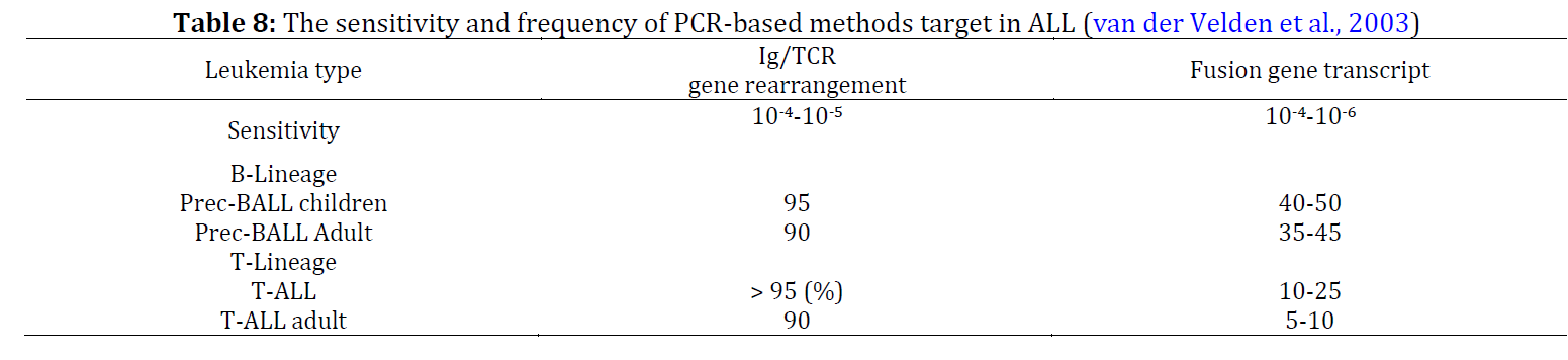
- van der Velden VHJ, Hochhaus A, Cazzaniga G, Szczepanski T, Gabert J, and Van Dongen JJM (2003). Detection of minimal residual disease in hematologic malignancies by real-time quantitative PCR: Principles, approaches, and laboratory aspects. Leukemia, 17(6): 1013-1034. https://doi.org/10.1038/sj.leu.2402922 [Google Scholar] PMid:12764363
- Vellichirammal NN, Chaturvedi NK, Joshi SS, Coulter DW, and Guda C (2021). Fusion genes as biomarkers in pediatric cancers: A review of the current state and applicability in diagnostics and personalized therapy. Cancer Letters, 499: 24-38. https://doi.org/10.1016/j.canlet.2020.11.015 [Google Scholar] PMid:33248210 PMCid:PMC9275405
- Waanders E, Gu Z, Dobson SM, Antić Ž, Crawford JC, and Ma X, Edmonson MN, Payne-Turner D, and van de Vorst M et al. (2020). Mutational landscape and patterns of clonal evolution in relapsed pediatric acute lymphoblastic leukemia. Blood Cancer Discovery, 1(1): 96-111. https://doi.org/10.1158/0008-5472.BCD-19-0041 [Google Scholar] PMid:32793890 PMCid:PMC7418874
- Weber A, Taube S, Zur Stadt U, Horstmann M, Krohn K, Bradtke J, and Christiansen H (2012). Quantification of minimal residual disease (MRD) in acute lymphoblastic leukemia (ALL) using amplicon-fusion-site polymerase chain reaction (AFS-PCR). Experimental Hematology and Oncology, 1: 33. https://doi.org/10.1186/2162-3619-1-33 [Google Scholar] PMid:23210797 PMCid:PMC3518178
- Weng XQ, Shen Y, Sheng Y, Chen B, Wang JH, Li JM, and Chen SJ (2013). Prognostic significance of monitoring leukemia-associated immunophenotypes by eight-color flow cytometry in adult B-acute lymphoblastic leukemia. Blood Cancer Journal, 3: e133. https://doi.org/10.1038/bcj.2013.31 [Google Scholar] PMid:23955588 PMCid:PMC3763385
- Willemse MJ, Seriu T, Hettinger K, d'Aniello E, Hop WC, Panzer-Grümayer ER, and van Dongen JJ (2002). Detection of minimal residual disease identifies differences in treatment response between T-ALL and precursor B-ALL. Blood: The Journal of the American Society of Hematology, 99(12): 4386-4393. https://doi.org/10.1182/blood.V99.12.4386 [Google Scholar] PMid:12036866
- Wu D, Sherwood A, Fromm JR, Winter SS, Dunsmore KP, Loh ML, and Robins H (2012). High-throughput sequencing detects minimal residual disease in acute T lymphoblastic leukemia. Science Translational Medicine, 4(134): 134ra63. https://doi.org/10.1126/scitranslmed.3003656 [Google Scholar]
- Yin JAL, O'Brien MA, Hills RK, Daly SB, Wheatley K, and Burnett AK (2012). Minimal residual disease monitoring by quantitative RT-PCR in core binding factor AML allows risk stratification and predicts relapse: Results of the United Kingdom MRC AML-15 trial. Blood: The Journal of the American Society of Hematology, 120(14): 2826-2835. https://doi.org/10.1182/blood-2012-06-435669 [Google Scholar] PMid:22875911
- Zhang L, Li Q, Li W, Liu B, Wang Y, Lin D, and Mi Y (2013a). Monitoring of minimal residual disease in acute myeloid leukemia with t (8; 21)(q22; q22). International Journal of Hematology, 97: 786-792. https://doi.org/10.1007/s12185-013-1344-6 [Google Scholar] PMid:23613269
- Zhang R, Liao J, Li G, Sun HQ, Shi YJ, and Yang JY (2013b). Real-time quantitative detection of E2A-PBX1 fusion gene in children with acute lymphoblastic leukemia and its clinical application in minimal residual disease monitoring. Zhongguo Dang Dai Er Ke Za Zhi [Chinese Journal of Contemporary Pediatrics], 15(6): 440-443. [Google Scholar] PMid:23791058