
International
ADVANCED AND APPLIED SCIENCES
EISSN: 2313-3724, Print ISSN: 2313-626X
Frequency: 12
![]()
Volume 10, Issue 12 (December 2023), Pages: 29-41

----------------------------------------------
Review Paper
Current trends of artificial intelligence and applications in digital pathology: A comprehensive review
Author(s):
Affiliation(s):
1Department of Biomedical Engineering, Manipal Institute of Technology (MIT), Manipal Academy of Higher Education, Manipal, India
2Department of Computer Science and Engineering, Manipal Institute of Technology (MIT), Manipal Academy of Higher Education, Manipal, India
3Prasanna School of Public Health, Manipal Academy of Higher Education, Manipal, India
Full text

* Corresponding Author.
 Corresponding author's ORCID profile: https://orcid.org/0000-0002-3345-360X
Digital Object Identifier (DOI)
https://doi.org/10.21833/ijaas.2023.12.004
Abstract
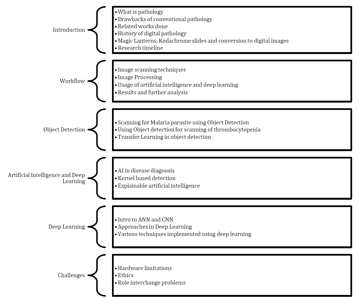
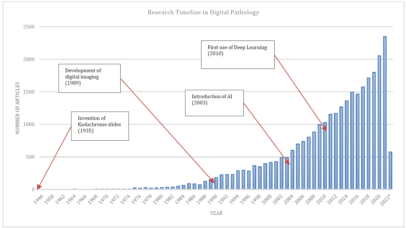
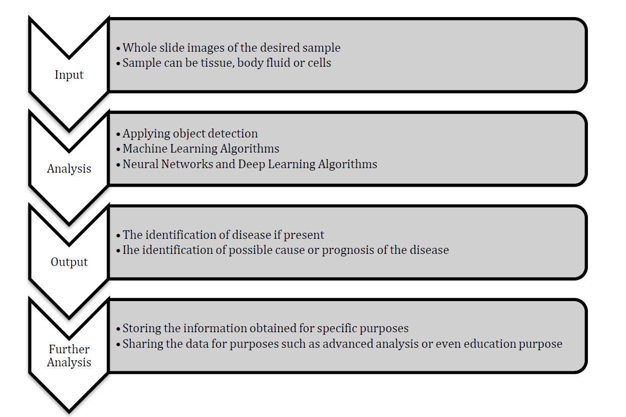
Digital pathology is a field that blends various techniques for obtaining, analyzing, sharing, and saving information about pathology. This information often comes from digitized microscope slides. Digital pathology also uses artificial intelligence (AI) to help reduce errors made by humans. This review talks about digital pathology and the new techniques linked to it. Instead of traditional microscopes, digital pathology employs virtual microscopy and whole-slide imaging. It marks a major improvement over old pathology methods, which had several problems. Digital methods use computers and machines to solve these issues. The basic process of digital pathology has three parts: the input stage, the analysis stage, and the output stage, which includes storing the information. This review focuses on two main techniques: object detection and its smaller methods, and the use of AI and its specific approaches like explainable AI (XAI) and deep learning. The paper also discusses various deep learning methods, mainly used to detect different types of cancer. It also acknowledges that not every method is perfect, so we discuss various challenges and limitations of digital pathology techniques that need to be solved before these methods can be widely used.
© 2023 The Authors. Published by IASE.
This is an
Keywords
Artificial intelligence, Digital pathology, Object detection, Digital health
Article history
Received 1 July 2023, Received in revised form 11 October 2023, Accepted 9 November 2023
Acknowledgment
No Acknowledgment.
Compliance with ethical standards
Conflict of interest: The author(s) declared no potential conflicts of interest with respect to the research, authorship, and/or publication of this article.
Citation:
Goswami NG, Karnad S, Sampathila N, Bairy GM, Chadaga K, and Swathi KS (2023). Current trends of artificial intelligence and applications in digital pathology: A comprehensive review. International Journal of Advanced and Applied Sciences, 10(12): 29-41
Figures
Fig. 1 Fig. 2 Fig. 3 Fig. 4 Fig. 5 Fig. 6 Fig. 7 Fig. 8 Fig. 9
Tables
Table 1 Table 2 Table 3 Table 4
----------------------------------------------
References (72)
- Aeffner F, Zarella MD, Buchbinder N, Bui MM, Goodman MR, Hartman DJ, and Bowman D (2019). Introduction to digital image analysis in whole-slide imaging: a white paper from the digital pathology association. Journal of Pathology Informatics, 10(1): 9. https://doi.org/10.4103/jpi.jpi_82_18 [Google Scholar] PMid:30984469 PMCid:PMC6437786
- Alake R (2022). Deep learning: GoogLeNet explained: One of the initial convolutional neural network that dared to go deeper. Available online at: https://towardsdatascience.com/deep-learning-googlenet-explained-de8861c82765
- Armstrong N and Hilton P (2014). Doing diagnosis: Whether and how clinicians use a diagnostic tool of uncertain clinical utility. Social Science and Medicine, 120: 208-214. https://doi.org/10.1016/j.socscimed.2014.09.032 [Google Scholar] PMid:25259659
- Barabási AL, Gulbahce N, and Loscalzo J (2011). Network medicine: A network-based approach to human disease. Nature Reviews Genetics, 12(1): 56-68. https://doi.org/10.1038/nrg2918 [Google Scholar] PMid:21164525 PMCid:PMC3140052
- Beitzel SM, Jensen EC, and Frieder O (2009). Map. In: Liu L and Özsu MT (Eds.), Encyclopedia of database systems: 1691-1692. Springer US, Boston, USA. https://doi.org/10.1007/978-0-387-39940-9_492 [Google Scholar]
- BenTaieb A and Hamarneh G (2019). Deep learning models for digital pathology. ArXiv Preprint ArXiv: 1910.12329. https://doi.org/10.48550/arXiv.1910.12329 [Google Scholar]
- Bisong E and Bisong E (2019). Google colaboratory. In: Bisong E (Ed.), Building machine learning and deep learning models on Google cloud platform: 59-64. Apress, Berkeley, USA. https://doi.org/10.1007/978-1-4842-4470-8_7 [Google Scholar]
- Block DR and Algeciras-Schimnich A (2013). Body fluid analysis: clinical utility and applicability of published studies to guide interpretation of today’s laboratory testing in serous fluids. Critical Reviews in Clinical Laboratory Sciences, 50(4-5): 107-124. https://doi.org/10.3109/10408363.2013.844679 [Google Scholar] PMid:24156653
- Bochkovskiy A, Wang CY, and Liao HYM (2020). YOLOv4: Optimal speed and accuracy of object detection. ArXiv Preprint ArXiv: 2004.10934. https://doi.org/10.48550/arXiv.2004.10934 [Google Scholar]
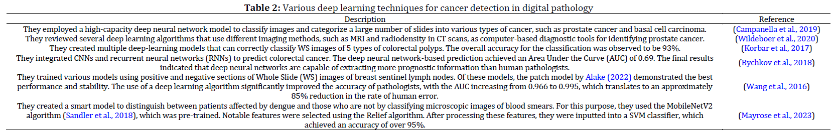
- Bychkov D, Linder N, Turkki R, Nordling S, Kovanen PE, Verrill C, and Lundin J (2018). Deep learning based tissue analysis predicts outcome in colorectal cancer. Scientific Reports, 8(1): 3395. https://doi.org/10.1038/s41598-018-21758-3 [Google Scholar] PMid:29467373 PMCid:PMC5821847
- Campanella G, Hanna MG, Geneslaw L, Miraflor A, Werneck Krauss Silva V, Busam KJ, and Fuchs TJ (2019). Clinical-grade computational pathology using weakly supervised deep learning on whole slide images. Nature Medicine, 25(8): 1301-1309. https://doi.org/10.1038/s41591-019-0508-1 [Google Scholar] PMid:31308507 PMCid:PMC7418463
- Cao JS, Lu ZY, Chen MY, Zhang B, Juengpanich S, Hu JH, and Cai XJ (2021). Artificial intelligence in gastroenterology and hepatology: Status and challenges. World Journal of Gastroenterology, 27(16): 1664-1690. https://doi.org/10.3748/wjg.v27.i16.1664 [Google Scholar] PMid:33967550 PMCid:PMC8072192
- Chico V (2018). The impact of the general data protection regulation on health research. British Medical Bulletin, 128(1): 109-118. https://doi.org/10.1093/bmb/ldy038 [Google Scholar] PMid:30445448
- Deng S, Zhang X, Yan W, Chang EIC, Fan Y, Lai M, and Xu Y (2020). Deep learning in digital pathology image analysis: A survey. Frontiers of Medicine, 14: 470-487. https://doi.org/10.1007/s11684-020-0782-9 [Google Scholar] PMid:32728875
- Došilović FK, Brčić M, and Hlupić N (2018). Explainable artificial intelligence: A survey. In the 2018 41st International Convention on Information and Communication Technology, Electronics And Microelectronics, IEEE, Opatija, Croatia: 0210-0215. https://doi.org/10.23919/MIPRO.2018.8400040 [Google Scholar]
- Evans T, Retzlaff CO, Geißler C, Kargl M, Plass M, and Müller H, and Holzinger A (2022). The explainability paradox: Challenges for xAI in digital pathology. Future Generation Computer Systems, 133: 281-296. https://doi.org/10.1016/j.future.2022.03.009 [Google Scholar]
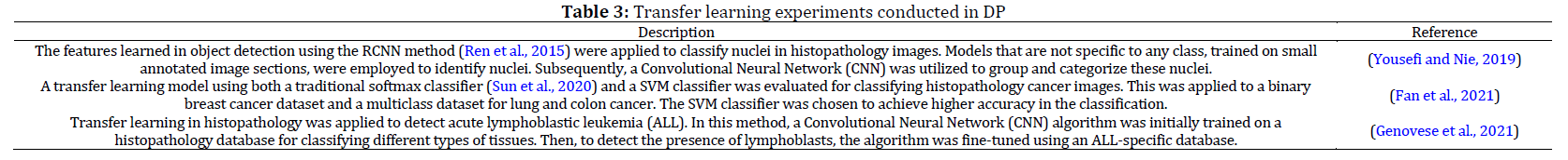
- Fan J, Lee J, and Lee Y (2021). A transfer learning architecture based on a support vector machine for histopathology image classification. Applied Sciences, 11(14): 6380. https://doi.org/10.3390/app11146380 [Google Scholar]
- Farahani N, Parwani AV, and Pantanowitz L (2015). Whole slide imaging in pathology: Advantages, limitations, and emerging perspectives. Pathology and Laboratory Medicine International, 7: 23-33. https://doi.org/10.2147/PLMI.S59826 [Google Scholar]
- Genovese A, Hosseini MS, Piuri V, Plataniotis KN, and Scotti F (2021). Histopathological transfer learning for Acute Lymphoblastic Leukemia detection. In the 2021 IEEE International Conference on Computational Intelligence and Virtual Environments for Measurement Systems and Applications, IEEE, Hong Kong, China: 1-6. https://doi.org/10.1109/CIVEMSA52099.2021.9493677 [Google Scholar]
- Gilmore H, Feldman M, Madabhushi A, Janowczyk A, and Zuo R (2019). HistoQC: An open-source quality control tool for digital pathology slides. JCO Clinical Cancer Informatics, 3: 1-7. https://doi.org/10.1200/CCI.18.00157 [Google Scholar] PMid:30990737 PMCid:PMC6552675
- Goebel R, Chander A, Holzinger K, Lecue F, Akata Z, Stumpf S, and Holzinger A (2018). Explainable AI: The new 42? In the Machine Learning and Knowledge Extraction: Second IFIP TC 5, TC 8/WG 8.4, 8.9, TC 12/WG 12.9 International Cross-Domain Conference, CD-MAKE 2018, Springer International Publishing, Hamburg, Germany, 295-303. https://doi.org/10.1007/978-3-319-99740-7_21 [Google Scholar]
- He Y, Zhao H, and Wong ST (2021). Deep learning powers cancer diagnosis in digital pathology. Computerized Medical Imaging and Graphics, 88: 101820. https://doi.org/10.1016/j.compmedimag.2020.101820 [Google Scholar] PMid:33453648 PMCid:PMC7902448
- Hegde N, Hipp JD, Liu Y, Emmert-Buck M, Reif E, Smilkov D, and Stumpe MC (2019). Similar image search for histopathology: SMILY. NPJ Digital Medicine, 2(1): 56. https://doi.org/10.1038/s41746-019-0131-z [Google Scholar] PMid:31304402 PMCid:PMC6588631
- Hollensead SC, Lockwood WB, and Elin RJ (2004). Errors in pathology and laboratory medicine: consequences and prevention. Journal of Surgical Oncology, 88(3): 161-181. https://doi.org/10.1002/jso.20125 [Google Scholar] PMid:15562462
- Holzinger A, Langs G, Denk H, Zatloukal K, and Müller H (2019). Causability and explainability of artificial intelligence in medicine. Wiley Interdisciplinary Reviews: Data Mining and Knowledge Discovery, 9(4): e1312. https://doi.org/10.1002/widm.1312 [Google Scholar] PMid:32089788 PMCid:PMC7017860
- Hou L, Samaras D, Kurc TM, Gao Y, Davis JE, and Saltz JH (2016). Patch-based convolutional neural network for whole slide tissue image classification. In the Proceedings of the IEEE Conference on Computer Vision and Pattern Recognition: 2424-2433. https://doi.org/10.1109/CVPR.2016.266 [Google Scholar] PMid:27795661 PMCid:PMC5085270
- Hunt JL (2008). Identifying cross contaminants and specimen mix-ups in surgical pathology. Advances in Anatomic Pathology, 15(4): 211-217. https://doi.org/10.1097/PAP.0b013e31817bf596 [Google Scholar] PMid:18580097
- Imambi S, Prakash KB, and Kanagachidambaresan GR (2021). PyTorch. In: Prakash KB and Kanagachidambaresan GR (Eds.), Programming with tensorFlow. EAI/Springer Innovations in Communication and Computing. Springer, Cham, Switzerland. https://doi.org/10.1007/978-3-030-57077-4 [Google Scholar]
- Jha S and Topol EJ (2016). Adapting to artificial intelligence: Radiologists and pathologists as information specialists. JAMA, 316(22): 2353-2354. https://doi.org/10.1001/jama.2016.17438 [Google Scholar] PMid:27898975
- Khandekar R, Shastry P, Jaishankar S, Faust O, and Sampathila N (2021). Automated blast cell detection for Acute Lymphoblastic Leukemia diagnosis. Biomedical Signal Processing and Control, 68: 102690. https://doi.org/10.1016/j.bspc.2021.102690 [Google Scholar]
- Khosravi P, Kazemi E, Imielinski M, Elemento O, and Hajirasouliha I (2018). Deep convolutional neural networks enable discrimination of heterogeneous digital pathology images. EBioMedicine, 27: 317-328. https://doi.org/10.1016/j.ebiom.2017.12.026 [Google Scholar] PMid:29292031 PMCid:PMC5828543
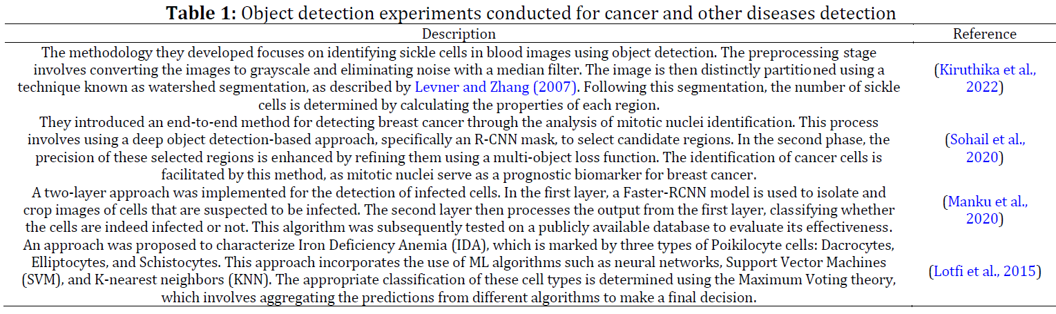
- Kiruthika V, Vallikannu AL, and Vimalarani G (2021). Automatic sickle cell Anaemia detection using image processing technique. In the International Conference on Micro-Electronics and Telecommunication Engineering, Springer Nature, Singapore, Singapore: 281-288. https://doi.org/10.1007/978-981-16-8721-1_27 [Google Scholar]
- Korbar B, Olofson AM, Miraflor AP, Nicka CM, Suriawinata MA, Torresani L, and Hassanpour S (2017). Deep learning for classification of colorectal polyps on whole-slide images. Journal of Pathology Informatics, 8(1): 30. https://doi.org/10.4103/jpi.jpi_34_17 [Google Scholar] PMid:28828201 PMCid:PMC5545773
- Krishnadas P and Sampathila N (2021). Automated detection of malaria implemented by deep learning in PyTorch. In the 2021 IEEE International Conference on Electronics, Computing and Communication Technologies, IEEE, Bangalore, India: 1-5. https://doi.org/10.1109/CONECCT52877.2021.9622608 [Google Scholar] PMid:34075874
- Kuc RB (1979). Application of Kalman filtering techniques to diagnostic ultrasound. Ultrasonic Imaging, 1(2): 105-120. https://doi.org/10.1016/0161-7346(79)90010-5 [Google Scholar] PMid:575816
- Levner I and Zhang H (2007). Classification-driven watershed segmentation. IEEE Transactions on Image Processing, 16(5): 1437-1445. https://doi.org/10.1109/TIP.2007.894239 [Google Scholar] PMid:17491471
- Liu J, Wu J, Sun L, and Zhu H (2020). Image data model optimization method based on cloud computing. Journal of Cloud Computing, 9: 1-10. https://doi.org/10.1186/s13677-020-00178-7 [Google Scholar]
- Lotfi M, Nazari B, Sadri S, and Sichani NK (2015). The detection of dacrocyte, schistocyte and elliptocyte cells in iron deficiency anemia. In the 2015 2nd International Conference on Pattern Recognition and Image Analysis, IEEE, Rasht, Iran: 1-5. https://doi.org/10.1109/PRIA.2015.7161628 [Google Scholar] PMid:27252956 PMCid:PMC4623536
- Madabhushi A and Lee G (2016). Image analysis and machine learning in digital pathology: Challenges and opportunities. Medical Image Analysis, 33: 170-175. https://doi.org/10.1016/j.media.2016.06.037 [Google Scholar] PMid:27423409 PMCid:PMC5556681
- Majno G and Joris I (1973). The microscope in the history of pathology with a note on the pathology of fat cells. Virchows Archiv A, 360: 273-286. https://doi.org/10.1007/BF00548349 [Google Scholar] PMid:4201096
- Manku RR, Sharma A, and Panchbhai A (2020). Malaria detection and classification. ArXiv Preprint ArXiv:2011.14329. https://doi.org/10.48550/arXiv.2011.14329 [Google Scholar]
- Marée R, Dallongeville S, Olivo-Marin JC, and Meas-Yedid V (2016). An approach for detection of glomeruli in multisite digital pathology. In the 2016 IEEE 13th International Symposium on Biomedical Imaging, IEEE, Prague, Czech Republic: 1033-1036. https://doi.org/10.1109/ISBI.2016.7493442 [Google Scholar]
- Mayrose H, Niranjana S, Bairy GM, Edwankar H, Belurkar S, and Saravu K (2021). Computer vision approach for the detection of thrombocytopenia from microscopic blood smear images. In the 2021 IEEE International Conference on Electronics, Computing and Communication Technologies, IEEE, Bangalore, India: 1-5. https://doi.org/10.1109/CONECCT52877.2021.9622688 [Google Scholar]
- Mayrose H, Sampathila N, Bairy GM, Belurkar S, Saravu K, Basu A, and Khan S (2023). Intelligent algorithm for detection of dengue using mobilenetv2‐based deep features with lymphocyte nucleus. Expert Systems, 40(4): e12904. https://doi.org/10.1111/exsy.12904 [Google Scholar]
- Merino A, Puigví L, Boldú L, Alférez S, and Rodellar J (2018). Optimizing morphology through blood cell image analysis. International Journal of Laboratory Hematology, 40: 54-61. https://doi.org/10.1111/ijlh.12832 [Google Scholar] PMid:29741256
- Morrison AO and Gardner JM (2015). Microscopic image photography techniques of the past, present, and future. Archives of Pathology and Laboratory Medicine, 139(12): 1558-1564. https://doi.org/10.5858/arpa.2014-0315-RA [Google Scholar] PMid:25989285
- Mouliou DS and Gourgoulianis KI (2021). False-positive and false-negative COVID-19 cases: Respiratory prevention and management strategies, vaccination, and further perspectives. Expert Review of Respiratory Medicine, 15(8): 993-1002. https://doi.org/10.1080/17476348.2021.1917389 [Google Scholar] PMid:33896332 PMCid:PMC8074645
- Mukti IZ and Biswas D (2019). Transfer learning based plant diseases detection using ResNet50. In the 2019 4th International conference on electrical information and communication technology, IEEE, Khulna, Bangladesh: 1-6. https://doi.org/10.1109/EICT48899.2019.9068805 [Google Scholar]
- Nakamura Y (2019). The role and necessity of sentinel lymph node biopsy for invasive melanoma. Frontiers in Medicine, 6: 231. https://doi.org/10.3389/fmed.2019.00231 [Google Scholar] PMid:31696119 PMCid:PMC6817613
- Niazi MKK, Parwani AV, and Gurcan MN (2019). Digital pathology and artificial intelligence. The Lancet Oncology, 20(5): e253-e261. https://doi.org/10.1016/S1470-2045(19)30154-8 [Google Scholar] PMid:31044723
- Orlov N, Shamir L, Macura T, Johnston J, Eckley DM, and Goldberg IG (2008). WND-CHARM: Multi-purpose image classification using compound image transforms. Pattern Recognition Letters, 29(11): 1684-1693. https://doi.org/10.1016/j.patrec.2008.04.013 [Google Scholar] PMid:18958301 PMCid:PMC2573471
- Pantanowitz L, Valenstein PN, Evans AJ, Kaplan KJ, Pfeifer JD, Wilbur DC, and Colgan TJ (2011). Review of the current state of whole slide imaging in pathology. Journal of Pathology Informatics, 2(1): 36. https://doi.org/10.4103/2153-3539.83746 [Google Scholar] PMid:21886892 PMCid:PMC3162745
- Park MK (2008). Pathophysiology. In: Myung K and Park RG (Eds.), Pediatric cardiology for practitioners. Elsevier, Philadelphia, USA. https://doi.org/10.1016/B978-0-323-04636-7.50014-9 [Google Scholar]
- Pattanaik PA, Swarnkar T, and Sheet D (2017). Object detection technique for malaria parasite in thin blood smear images. In the 2017 IEEE International Conference on Bioinformatics and Biomedicine, IEEE, Kansas City, USA: 2120-2123. https://doi.org/10.1109/BIBM.2017.8217986 [Google Scholar]
- Ren S, He K, Girshick R, and Sun J (2015). Faster R-CNN: Towards real-time object detection with region proposal networks. In the Proceedings of Advances in Neural Information Processing Systems 28 (NIPS 2015): 91-99. Available online at: https://proceedings.neurips.cc/paper_files/paper/2015 [Google Scholar]
- Ren Z, Lam EY, and Zhao J (2021). Learning-based cell detection in digital pathology. In the Conference on Lasers and Electro-Optics (CLEO), QELS_Fundamental Science, Optica Publishing Group, San Jose, USA. https://doi.org/10.1364/CLEO_AT.2021.JW1A.184 [Google Scholar] PMid:34717623 PMCid:PMC8557613
- Salto-Tellez M, Maxwell P, and Hamilton P (2019). Artificial intelligence-the third revolution in pathology. Histopathology, 74(3): 372-376. https://doi.org/10.1111/his.13760 [Google Scholar] PMid:30270453
- Sandler M, Howard A, Zhu M, Zhmoginov A, and Chen LC (2018). Mobilenetv2: Inverted residuals and linear bottlenecks. In the Proceedings of the IEEE Conference on Computer Vision and Pattern Recognition, Salt Lake City, USA: 4510-4520. https://doi.org/10.1109/CVPR.2018.00474 [Google Scholar]
- Senaras C, Niazi MKK, Lozanski G, and Gurcan MN (2018). DeepFocus: Detection of out-of-focus regions in whole slide digital images using deep learning. PLOS ONE, 13(10): e0205387. https://doi.org/10.1371/journal.pone.0205387 [Google Scholar] PMid:30359393 PMCid:PMC6201886
- Shet NR and Sampathila N (2015). An image processing approach for screening of malaria. In the Proceedings of NJCIET 2015, Canara Engineering College, Mangalore, India: 395-399. [Google Scholar]
- Sohail A, Mukhtar MA, Khan A, Zafar MM, Zameer A, and Khan S (2020). Deep object detection based mitosis analysis in breast cancer histopathological images. Arxiv Preprint Arxiv: 2003.08803. https://doi.org/10.48550/arXiv.2003.08803 [Google Scholar]
- Solano-Rojas B, Villalón-Fonseca R, and Marín-Raventós G (2020). Alzheimer’s disease early detection using a low cost three-dimensional densenet-121 architecture. In the Impact of Digital Technologies on Public Health in Developed and Developing Countries: 18th International Conference, Springer International Publishing, Hammamet, Tunisia: 3-15. https://doi.org/10.1007/978-3-030-51517-1_1 [Google Scholar] PMCid:PMC7313295
- Stoeklé HC, Mamzer-Bruneel MF, Frouart CH, Le Tourneau C, Laurent-Puig P, Vogt G, and Hervé C (2018). Molecular tumor boards: Ethical issues in the new era of data medicine. Science and Engineering Ethics, 24: 307-322. https://doi.org/10.1007/s11948-017-9880-8 [Google Scholar] PMid:28281147 PMCid:PMC5799317
- Sun L, Li W, Ning X, Zhang L, Dong X, and He W (2020). Gradient-enhanced softmax for face recognition. IEICE Transactions on Information and Systems, 103(5): 1185-1189. https://doi.org/10.1587/transinf.2019EDL8103 [Google Scholar]
- Suthaharan S (2016). Support vector machine. In: Suthaharan S (Ed.), Machine learning models and algorithms for big data classification: Thinking with examples for effective learning: 207-235. Springer, Boston, USA. https://doi.org/10.1007/978-1-4899-7641-3_9 [Google Scholar]
- Sze V, Chen YH, Emer J, Suleiman A, and Zhang Z (2017). Hardware for machine learning: Challenges and opportunities. In the 2017 IEEE Custom Integrated Circuits Conference, IEEE, Austin, USA: 1-8. https://doi.org/10.1109/CICC.2017.7993626 [Google Scholar] PMCid:PMC5209811
- Trotter MJ, Larsen ET, Tait N, and Wright JJR (2009). Time study of clinical and nonclinical workload in pathology and laboratory medicine. American Journal of Clinical Pathology, 131(6): 759-767. https://doi.org/10.1309/AJCP8SKO6BUJQXHD [Google Scholar] PMid:19461078
- Wang D, Khosla A, Gargeya R, Irshad H, and Beck AH (2016). Deep learning for identifying metastatic breast cancer. Arxiv Preprint Arxiv: 1606.05718. https://doi.org/10.48550/arXiv.1606.05718 [Google Scholar]
- Wildeboer RR, van Sloun RJ, Wijkstra H, and Mischi M (2020). Artificial intelligence in multiparametric prostate cancer imaging with focus on deep-learning methods. Computer Methods and Programs in Biomedicine, 189: 105316. https://doi.org/10.1016/j.cmpb.2020.105316 [Google Scholar] PMid:31951873
- Wright JJ (2018). Historical insights for early adopters of whole slide imaging. Archives of Pathology and Laboratory Medicine, 142(2): 161-162. https://doi.org/10.5858/arpa.2017-0326-LE [Google Scholar] PMid:29372849
- Yilmaz A, Javed O, and Shah M (2006). Object tracking. ACM Computing Surveys, 38(4): 13. https://doi.org/10.1145/1177352.1177355 [Google Scholar]
- Yousefi S and Nie Y (2019). Transfer learning from nucleus detection to classification in histopathology images. In the 2019 IEEE 16th International Symposium on Biomedical Imaging, IEEE, Venice, Italy: 957-960. https://doi.org/10.1109/ISBI.2019.8759469 [Google Scholar] PMid:30686965 PMCid:PMC6336923