
International
ADVANCED AND APPLIED SCIENCES
EISSN: 2313-3724, Print ISSN: 2313-626X
Frequency: 12
![]()
Volume 9, Issue 4 (April 2022), Pages: 126-128

----------------------------------------------
Case Report
Title: A rare case of unilateral acute posterior multifocal placoid pigment epitheliopathy with features of Vogt-Koyanagi-Harada disease
Author(s): Pradeep Kumar Panigrahi *, Lipika Mehra, Alpana Mishra, Deergha Pareek
Affiliation(s):
Department of Ophthalmology, Institute of Medical Sciences and SUM Hospital, Siksha O Anusandhan (Deemed to be University), Bhubaneswar, India

* Corresponding Author.
 Corresponding author's ORCID profile: https://orcid.org/0000-0003-1236-2845
Digital Object Identifier:
https://doi.org/10.21833/ijaas.2022.04.015
Abstract:
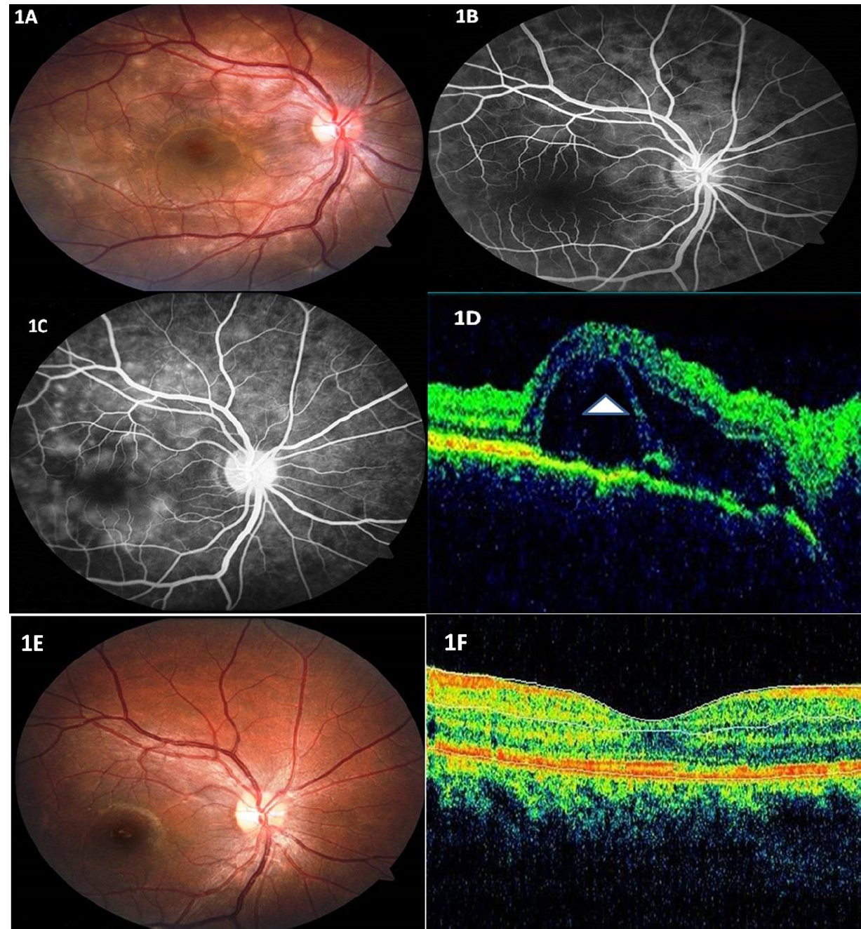
The purpose of this study is to report a rare case of unilateral acute posterior multifocal placoid pigment epitheliopathy (APMPPE) with features of Vogt-Koyanagi-Harada (VKH) disease. Both the diseases have their own unique presentations and can lead to vision loss in the affected eye. Overlapping features of both diseases in the same eye is rare. A 28-year-old healthy female presented with loss of vision in her right eye for 10 days duration. Visual acuity in the affected eye was 20/200, N36. Fundus examination revealed numerous creamy yellow lesions overlying the posterior pole with subretinal fluid. Further investigations including fundus fluorescein angiography and optical coherence tomography were suggestive of overlapping features of both APMPPE and VKH disease. Baseline laboratory investigations and markers for Sarcoidosis, Tuberculosis, and Syphilis were normal. The patient was treated with tapering doses of oral steroids. One month following initiation of treatment, the vision had improved to 20/20, N6 with complete resolution of subretinal fluid in the right eye. The overlapping clinical and imaging features suggest that both diseases may be a part of a common inflammatory process that secondarily damages the outer retinal structures.
© 2022 The Authors. Published by IASE.
This is an
Keywords: Acute posterior multifocal placoid, pigment epitheliopathy, Vogt-Koyanagi-Harada disease, Unilateral, Fundus fluorescein angiography, Optical coherence tomography
Article History: Received 18 November 2021, Received in revised form 12 February 2022, Accepted 14 February 2022
Acknowledgment
No Acknowledgment.
Compliance with ethical standards
Conflict of interest: The author(s) declared no potential conflicts of interest with respect to the research, authorship, and/or publication of this article.
Citation:
Panigrahi PK, Mehra L, and Mishra A et al. (2022). A rare case of unilateral acute posterior multifocal placoid pigment epitheliopathy with features of Vogt-Koyanagi-Harada disease. International Journal of Advanced and Applied Sciences, 9(4): 126-128
Figures
Tables
No Table
----------------------------------------------
References (4)
- Gass JDM (1968). Acute posterior multifocal placoid pigment epitheliopathy. Archives of Ophthalmology, 80(2): 177-185. https://doi.org/10.1001/archopht.1968.00980050179005 [Google Scholar] PMid:5661882
- Li B, Bentham RJ, and Gonder JR (2016). A case of unilateral and spontaneously resolving posterior uveitis with overlapping features of Vogt–Koyanagi–Harada disease and Acute Posterior Multifocal Placoid Pigment Epitheliopathy. Springerplus, 5(1): 1471. https://doi.org/10.1186/s40064-016-3132-2 [Google Scholar] PMid:27652046 PMCid:PMC5009056
- Moorthy RS, Inomata H, and Rao NA (1995). Vogt-Koyanagi-Harada syndrome. Survey of Ophthalmology, 39(4): 265-292. https://doi.org/10.1016/S0039-6257(05)80105-5 [Google Scholar]
- Vedantham V and Ramasamy K (2006). Atypical manifestations of acute posterior multifocal placoid pigment epitheliopathy. Indian Journal of Ophthalmology, 54(1): 49-52. https://doi.org/10.4103/0301-4738.21618 [Google Scholar] PMid:16531674