
International
ADVANCED AND APPLIED SCIENCES
EISSN: 2313-3724, Print ISSN: 2313-626X
Frequency: 12
![]()
Volume 9, Issue 4 (April 2022), Pages: 44-52

----------------------------------------------
Original Research Paper
Title: Deep transfer learning CNN based approach for COVID-19 detection
Author(s): Wazir Muhammad 1, *, Zuhaibuddin Bhutto 2, Syed Ali Raza Shah 3, Jalal Shah 2, Murtaza Hussain Shaikh 4, Ayaz Hussain 1, Imdadullah Thaheem 5, Shamshad Ali 1
Affiliation(s):
1Department of Electrical Engineering, Balochistan University of Engineering and Technology, Khuzdar, Pakistan
2Department of Computer System Engineering, Balochistan University of Engineering and Technology, Khuzdar, Pakistan
3Department of Mechanical Engineering, Balochistan University of Engineering and Technology, Khuzdar, Pakistan
4Department of Information Systems, Kyungsung University, Busan, South Korea
5Department of Energy Systems Engineering, Balochistan University of Engineering and Technology, Khuzdar, Pakistan

* Corresponding Author.
 Corresponding author's ORCID profile: https://orcid.org/0000-0002-3860-2213
Digital Object Identifier:
https://doi.org/10.21833/ijaas.2022.04.006
Abstract:
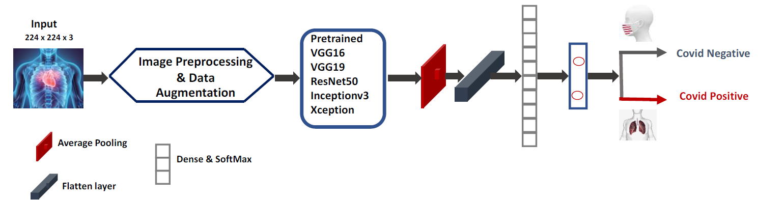
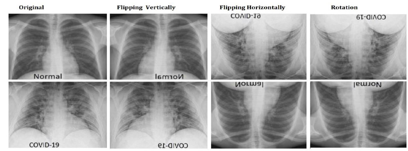
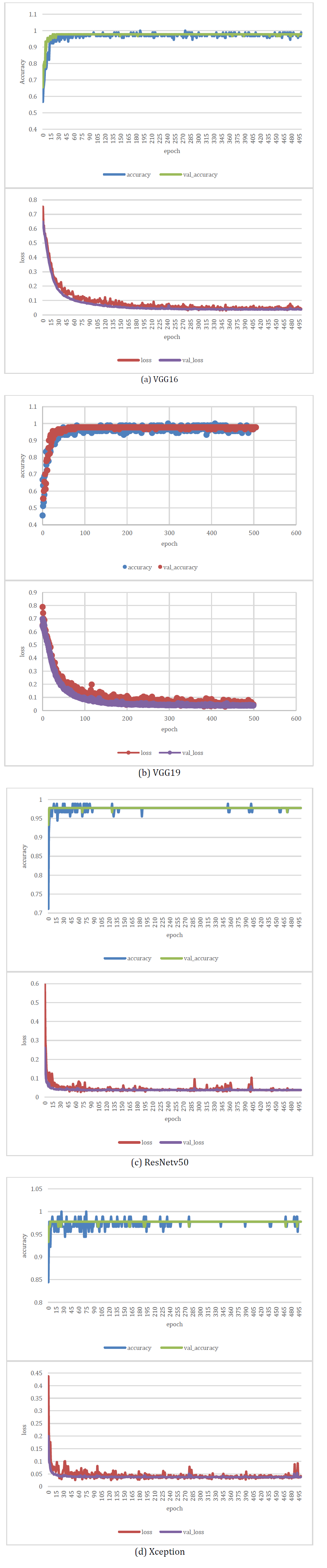
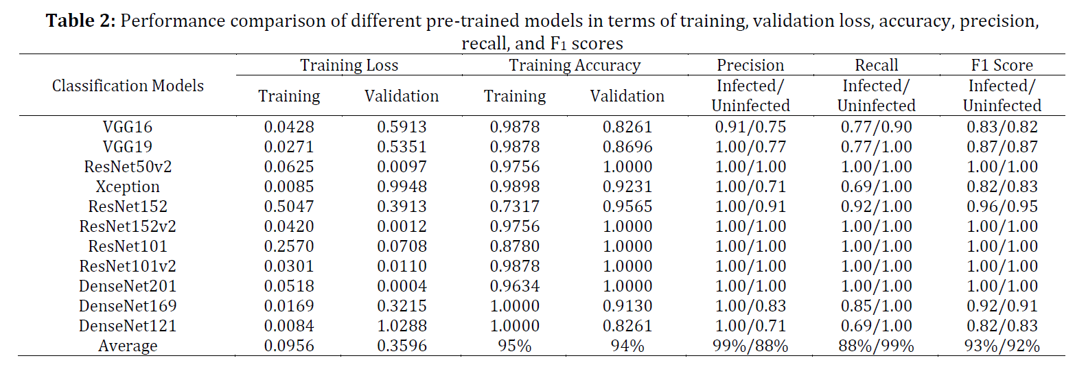
Recently, the novel coronavirus (Covid-19) and its different variants have spread rapidly across the world. Early-stage detection of COVID-19 is a challenging task due to the limited availability of Covid testing kits to the public. Conventionally, reverse transcription-polymerase chain reaction (RT-PCR) is the reliable test for the detection of COVID-19 which is time-consuming and costly. The aim of this work is to identify the COVID-19 symptoms with the help of a deep learning algorithm using chest X-Ray images. In order to improve the quality of chest X-Ray images, authors have further modified the pre-trained model with some extra CNN layers, such as the first layer is the average pooling layer and the other two are dense layers followed by ReLU with softmax activation function. The experimental results have been carried out on publicly available chest X-Ray images of COVID-19 to mark COVID-19 patients as positive and negative datasets. For evaluation purpose, we have used benchmark of pre-trained models such as VGG-16 (Visual Geometry Group), VGG19, Xception, ResNet152, ResNet152v2, ResNet101, ResNet101v2, DenseNet201, DenseNet169 and DenseNet121. On the benchmark datasets, viz. COVID-19 X-Ray images, an average improvement in terms of training/validation accuracy, precision, recall, and F1-scores scores were 95%, 94%, 99/88%, 99/88%, and 93/92% respectively. The results provide sufficient evidence that deep learning can be used efficiently for the detection of COVID-19 symptoms.
© 2022 The Authors. Published by IASE.
This is an
Keywords: COVID-19, Deep learning, Transfer learning, Chest X-ray
Article History: Received 20 September 2021, Received in revised form 31 January 2022, Accepted 7 February 2022
Acknowledgment
This work was supported by the Baluchistan University of Engineering and Technology, Khuzdar.
Compliance with ethical standards
Conflict of interest: The author(s) declared no potential conflicts of interest with respect to the research, authorship, and/or publication of this article.
Citation:
Muhammad W, Bhutto Z, and Shah SAR et al. (2022). Deep transfer learning CNN based approach for COVID-19 detection. International Journal of Advanced and Applied Sciences, 9(4): 44-52
Figures
Tables
----------------------------------------------
References (40)
- Abbas A, Abdelsamea MM, and Gaber MM (2021). Classification of COVID-19 in chest X-ray images using DeTraC deep convolutional neural network. Applied Intelligence, 51(2): 854-864. https://doi.org/10.1007/s10489-020-01829-7 [Google Scholar] PMid:34764548 PMCid:PMC7474514
- Apostolopoulos ID and Mpesiana TA (2020). Covid-19: Automatic detection from x-ray images utilizing transfer learning with convolutional neural networks. Physical and Engineering Sciences in Medicine, 43(2): 635-640. https://doi.org/10.1007/s13246-020-00865-4 [Google Scholar] PMid:32524445 PMCid:PMC7118364
- Ayan E and Ünver HM (2019). Diagnosis of pneumonia from chest X-ray images using deep learning. In the 2019 Scientific Meeting on Electrical-Electronics and Biomedical Engineering and Computer Science, IEEE, Istanbul, Turkey: 1-5. https://doi.org/10.1109/EBBT.2019.8741582 [Google Scholar] PMid:30711875
- Chollet F (2017). Xception: Deep learning with depthwise separable convolutions. In the Conference on Computer Vision and Pattern Recognition, IEEE, Honolulu, USA: 1251-1258. https://doi.org/10.1109/CVPR.2017.195 [Google Scholar]
- Chowdhury ME, Rahman T, Khandakar A, Mazhar R, Kadir MA, Mahbub ZB, and Islam MT (2020). Can AI help in screening viral and COVID-19 pneumonia? IEEE Access, 8: 132665-132676. https://doi.org/10.1109/ACCESS.2020.3010287 [Google Scholar]
- Cohen JP, Morrison P, Dao L, Roth K, Duong TQ, and Ghassemi M (2020). Covid-19 image data collection: Prospective predictions are the future. arXiv:2006.11988. https://doi.org/10.48550/arXiv.2006.11988 [Google Scholar]
- Deng J, Dong W, Socher R, Li LJ, Li K, and Fei-Fei L (2009). ImageNet: A large-scale hierarchical image database. In the Conference on Computer Vision and Pattern Recognition, IEEE, Miami, USA: 248-255. https://doi.org/10.1109/CVPR.2009.5206848 [Google Scholar]
- Hall LO, Paul R, Goldgof DB, and Goldgof GM (2020). Finding covid-19 from chest x-rays using deep learning on a small dataset. arXiv:2004.02060v4. https://doi.org/10.36227/techrxiv.12083964.v2 [Google Scholar]
- He K, Zhang X, Ren S, and Sun J (2016). Deep residual learning for image recognition. In the Conference on Computer Vision and Pattern Recognition, IEEE, Las Vegas, USA: 770-778. https://doi.org/10.1109/CVPR.2016.90 [Google Scholar] PMid:26180094
- Hemdan EED, Shouman MA, and Karar ME (2020). COVIDX-Net: A framework of deep learning classifiers to diagnose covid-19 in x-ray images. arXiv:2003.11055v1. https://doi.org/10.48550/arXiv.2003.11055 [Google Scholar]
- Homayouni H, Ray I, Ghosh S, Gondalia S, and Kahn MG (2021). Anomaly detection in COVID-19 time-series data. Computer Science, 2(4): 1-17. https://doi.org/10.1007/s42979-021-00658-w [Google Scholar] PMid:34027432 PMCid:PMC8132285
- Huang G, Liu Z, Van Der Maaten L, and Weinberger KQ (2017). Densely connected convolutional networks. In the Conference on Computer Vision and Pattern Recognition, IEEE, Honolulu, USA: 4700-4708. https://doi.org/10.1109/CVPR.2017.243 [Google Scholar] PMCid:PMC5598342
- Ismael AM and Şengür A (2021). Deep learning approaches for COVID-19 detection based on chest X-ray images. Expert Systems with Applications, 164: 114054. https://doi.org/10.1016/j.eswa.2020.114054 [Google Scholar] PMid:33013005 PMCid:PMC7521412
- Jain R, Gupta M, Taneja S, and Hemanth DJ (2021). Deep learning-based detection and analysis of COVID-19 on chest X-ray images. Applied Intelligence, 51(3): 1690-1700. https://doi.org/10.1007/s10489-020-01902-1 [Google Scholar] PMid:34764553 PMCid:PMC7544769
- Kermany DS, Goldbaum M, Cai W, Valentim CC, Liang H, Baxter SL, and Zhang K (2018). Identifying medical diagnoses and treatable diseases by image-based deep learning. Cell, 172(5): 1122-1131. https://doi.org/10.1016/j.cell.2018.02.010 [Google Scholar] PMid:29474911
- Khan AI, Shah JL, and Bhat MM (2020). CoroNet: A deep neural network for detection and diagnosis of COVID-19 from chest x-ray images. Computer Methods and Programs in Biomedicine, 196: 105581. https://doi.org/10.1016/j.cmpb.2020.105581 [Google Scholar] PMid:32534344 PMCid:PMC7274128
- Krizhevsky A, Sutskever I, and Hinton GE (2012). ImageNet classification with deep convolutional neural networks. Advances in Neural Information Processing Systems, 25: 1097-1105. [Google Scholar]
- Mobiny A, Cicalese PA, Zare S, Yuan P, Abavisani M, Wu CC, and Van Nguyen H (2020). Radiologist-level covid-19 detection using CT scans with detail-oriented capsule networks. arXiv:2004.07407v1. https://doi.org/10.48550/arXiv.2004.07407 [Google Scholar]
- Muhammad W and Aramvith S (2019). Multi-scale inception based super-resolution using deep learning approach. Electronics, 8(8): 892. https://doi.org/10.3390/electronics8080892 [Google Scholar]
- Muhammad W, Aramvith S, and Onoye T (2021a). Multi-scale Xception based depthwise separable convolution for single image super-resolution. PLOS ONE, 16(8): e0249278. https://doi.org/10.1371/journal.pone.0249278 [Google Scholar] PMid:34424911 PMCid:PMC8382202
- Muhammad W, Bhutto Z, Ansari A, Memon ML, Kumar R, Hussain A, and Ali S (2021b). Multi-path deep CNN with residual inception network for single image super-resolution. Electronics, 10(16): 1979. https://doi.org/10.3390/electronics10161979 [Google Scholar]
- Muhammad W, Ullah I, and Ashfaq M (2020). An introduction to deep convolutional neural networks with Keras. In: Mahrishi M, Hiran KK, Meena G, and Sharma P (Eds.), Machine learning and deep learning in real-time applications: 231-272. IGI Global, Pennsylvania, USA. https://doi.org/10.4018/978-1-7998-3095-5.ch011 [Google Scholar]
- Narin A, Kaya C, and Pamuk Z (2021). Automatic detection of coronavirus disease (covid-19) using x-ray images and deep convolutional neural networks. Pattern Analysis and Applications, 24: 1207–1220. https://doi.org/10.1007/s10044-021-00984-y [Google Scholar] PMid:33994847 PMCid:PMC8106971
- Özcan T and Baştürk A (2019). Static image-based emotion recognition using convolutional neural network. In the 27th Signal Processing and Communications Applications Conference, IEEE, Sivas, Turkey: 1-4. https://doi.org/10.1109/SIU.2019.8806408 [Google Scholar]
- Peled S and Yeshurun Y (2001). Superresolution in MRI: Application to human white matter fiber tract visualization by diffusion tensor imaging. Magnetic Resonance in Medicine: An Official Journal of the International Society for Magnetic Resonance in Medicine, 45(1): 29-35. https://doi.org/10.1002/1522-2594(200101)45:1<29::AID-MRM1005>3.0.CO;2-Z [Google Scholar]
- Qian H, Mao Y, Xiang W, and Wang Z (2010). Recognition of human activities using SVM multi-class classifier. Pattern Recognition Letters, 31(2): 100-111. https://doi.org/10.1016/j.patrec.2009.09.019 [Google Scholar]
- Russakovsky O, Deng J, Su H, Krause J, Satheesh S, Ma S, and Fei-Fei L (2015). ImageNet large scale visual recognition challenge. International Journal of Computer Vision, 115(3): 211-252. https://doi.org/10.1007/s11263-015-0816-y [Google Scholar]
- Sethy PK and Behera SK (2020). Detection of coronavirus disease (covid-19) based on deep features. Preprints 2020, 2020030300. https://doi.org/10.20944/preprints202003.0300.v1 [Google Scholar]
- Shan F, Gao Y, Wang J, Shi W, Shi N, Han M, and Shi Y (2020). Lung infection quantification of COVID-19 in CT images with deep learning. arXiv:2003.04655v3. https://doi.org/10.48550/arXiv.2003.04655 [Google Scholar]
- Shi W, Caballero J, Ledig C, Zhuang X, Bai W, Bhatia K, and Rueckert D (2013). Cardiac image super-resolution with global correspondence using multi-atlas patch match. In the International Conference on Medical Image Computing and Computer-Assisted Intervention, Springer, Nagoya, Japan: 9-16. https://doi.org/10.1007/978-3-642-40760-4_2 [Google Scholar] PMid:24505738
- Simonyan K and Zisserman A (2014). Very deep convolutional networks for large-scale image recognition. In the ICLR 2015: 3rd International Conference on Learning Representations 2015, San Diego, USA: 1-14. [Google Scholar]
- Singhal T (2020). “Rationalization of empiric antibiotic therapy”–A move towards preventing emergence of resistant infections. The Indian Journal of Pediatrics, 87: 945–950. https://doi.org/10.1007/s12098-019-03144-7 [Google Scholar] PMid:31912460
- Stephen O, Sain M, Maduh UJ, and Jeong DU (2019). An efficient deep learning approach to pneumonia classification in healthcare. Journal of Healthcare Engineering, 2019: 4180949. https://doi.org/10.1155/2019/4180949 [Google Scholar] PMid:31049186 PMCid:PMC6458916
- Strzelecki A (2020). The second worldwide wave of interest in coronavirus since the COVID-19 outbreaks in South Korea, Italy and Iran: A google trends study. Brain, Behavior, and Immunity, 88: 950–951. https://doi.org/10.1016/j.bbi.2020.04.042 [Google Scholar] PMid:32311493 PMCid:PMC7165085
- Szegedy C, Vanhoucke V, Ioffe S, Shlens J, and Wojna Z (2016). Rethinking the inception architecture for computer vision. In the IEEE Conference on Computer Vision and Pattern Recognition, IEEE, Las Vegas, USA: 2818-2826. https://doi.org/10.1109/CVPR.2016.308 [Google Scholar]
- Tajbakhsh N, Shin JY, Gurudu SR, Hurst RT, Kendall CB, Gotway MB, and Liang J (2016). Convolutional neural networks for medical image analysis: Full training or fine tuning? IEEE Transactions on Medical Imaging, 35(5): 1299-1312. https://doi.org/10.1109/TMI.2016.2535302 [Google Scholar] PMid:26978662
- Varshni D, Thakral K, Agarwal L, Nijhawan R, and Mittal A (2019). Pneumonia detection using CNN based feature extraction. In the IEEE International Conference on Electrical, Computer and Communication Technologies, IEEE, Coimbatore, India: 1-7. https://doi.org/10.1109/ICECCT.2019.8869364 [Google Scholar]
- Wang L, Lin ZQ, and Wong A (2020a). Covid-net: A tailored deep convolutional neural network design for detection of covid-19 cases from chest x-ray images. Scientific Reports, 10(1): 1-12. https://doi.org/10.1038/s41598-020-76550-z [Google Scholar] PMid:33177550 PMCid:PMC7658227
- Wang W, Xu Y, Gao R, Lu R, Han K, Wu G, and Tan W (2020b). Detection of SARS-CoV-2 in different types of clinical specimens. JAMA, 323(18): 1843-1844. https://doi.org/10.1001/jama.2020.3786 [Google Scholar]
- Wu Z and McGoogan JM (2020). Characteristics of and important lessons from the coronavirus disease 2019 (COVID-19) outbreak in China: Summary of a report of 72 314 cases from the Chinese center for disease control and prevention. JAMA, 323(13): 1239-1242. https://doi.org/10.1001/jama.2020.2648 [Google Scholar] PMid:32091533