
International
ADVANCED AND APPLIED SCIENCES
EISSN: 2313-3724, Print ISSN: 2313-626X
Frequency: 12
![]()
Volume 9, Issue 12 (December 2022), Pages: 135-144

----------------------------------------------
Original Research Paper
Comparative study on early recognition and identifying diabetic retinopathy with different layers in CNN
Author(s): Gorli L. Aruna Kumari 1, *, Poosapati Padmaja 2, Jaya G. Suma 3
Affiliation(s):
1Department of CSE, Gitam School of Technology, Gitam Deemed to be University, Visakhapatnam, India
2Department of IT, Anil Neerukonda Institute of Technology and Science, Visakhapatnam, India
3Department of IT, College of Engineering, Jawaharlal Nehru Technological University, Kakinada, India

* Corresponding Author.
 Corresponding author's ORCID profile: https://orcid.org/0000-0002-8856-5465
Digital Object Identifier:
https://doi.org/10.21833/ijaas.2022.12.017
Abstract:
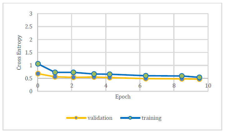
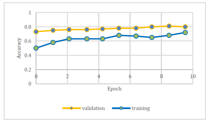
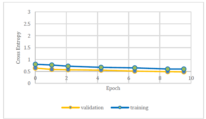
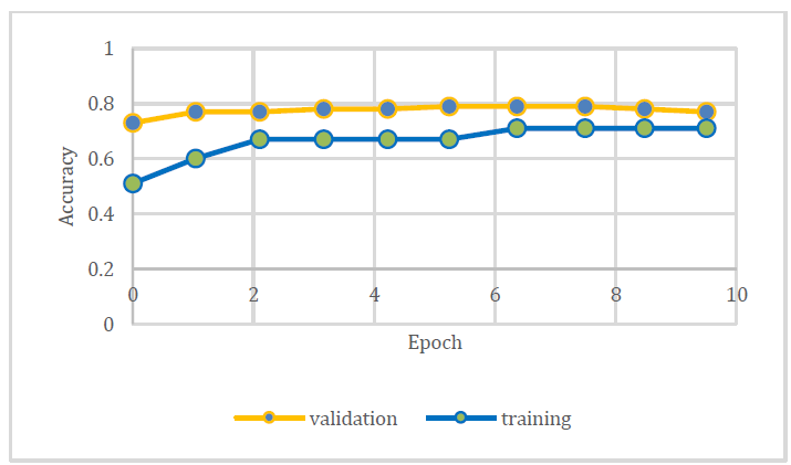
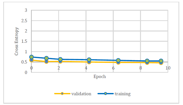
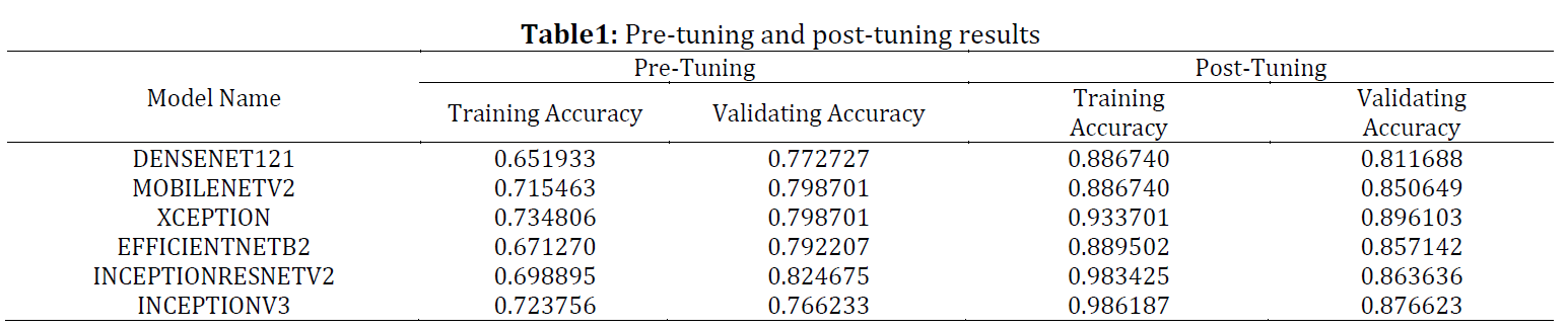
Diabetes is the most prevalent condition worldwide, and diabetic retinopathy (DR) is a subsequent condition caused by acute diabetic cases. It causes severe degeneration of the retina. The compounding blood vessels bloat and often burst, causing fluid leaks in the aqueous humor. This, in turn, causes the creation of undesirable nerve fiber infractions from the occlusion of arteries. Diagnosis requires a manual retinal examination that can often be inconsistent and deliberate with potential flaws in the diagnosis. Early detection through an ophthalmologist is paramount to prevent the prognosis of severe vision loss. Considering the current leap of machine learning in the field of healthcare, early detection of DR can be potentially made efficient with intelligent systems. This research proposes methodologies to fine-tune the existing pre-trained architectures, attaining the classification accuracies of 98% to classify the ocular fundus images which identify early prediction of diabetes. Additionally, this study presents an exposition of other equally scrutinized approaches to ultimately showcase a deep neural network architecture that can precisely classify normal fundus and degenerated fundus from the lowest to the most severe hierarchy. Among several layers in the CNN model pre-tuning and post-tuning exception layers outperformed with good results.
© 2022 The Authors. Published by IASE.
This is an
Keywords: Deep neural network, Classification, Convolution neural network, Data mining, Diabetic retinopathy
Article History: Received 10 March 2022, Received in revised form 9 June 2022, Accepted 7 September 2022
Acknowledgment
No Acknowledgment.
Compliance with ethical standards
Conflict of interest: The author(s) declared no potential conflicts of interest with respect to the research, authorship, and/or publication of this article.
Citation:
Kumari GLA, Padmaja P, and Suma JG (2022). Comparative study on early recognition and identifying diabetic retinopathy with different layers in CNN. International Journal of Advanced and Applied Sciences, 9(12): 135-144
Figures
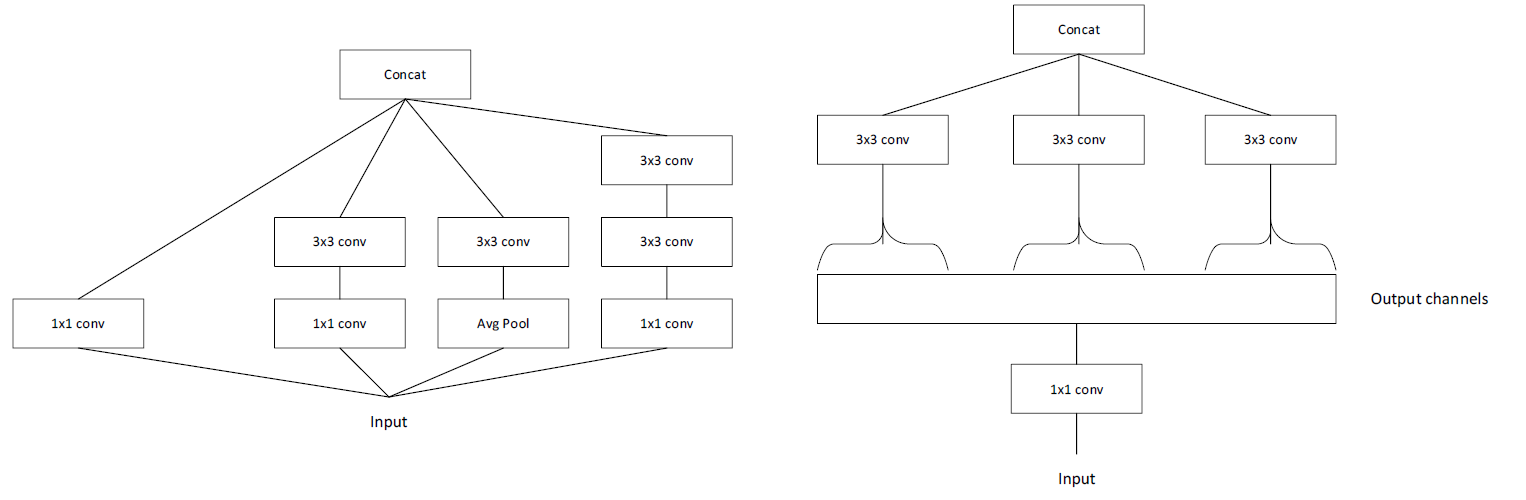
Fig. 1 Fig. 2 Fig. 3 Fig. 4 Fig. 5 Fig. 6 Fig. 7 Fig. 8 Fig. 9 Fig. 10 Fig. 11 Fig. 12 Fig. 13 Fig. 14 Fig. 15 Fig. 16 Fig. 17 Fig. 18 Fig. 19
Tables
----------------------------------------------
References (30)
- Acharya UR, Lim CM, Ng EYK, Chee C, and Tamura T (2009). Computer-based detection of diabetes retinopathy stages using digital fundus images. Proceedings of the Institution of Mechanical Engineers, part H: Journal of Engineering in Medicine, 223(5): 545-553. https://doi.org/10.1243/09544119JEIM486 [Google Scholar] PMid:19623908
- Amin J, Sharif M, and Yasmin M (2016). A review on recent developments for detection of diabetic retinopathy. Scientifica, 2016: 6838976. https://doi.org/10.1155/2016/6838976 [Google Scholar] PMid:27777811 PMCid:PMC5061953
- Anant KA, Ghorpade T, and Jethani V (2017). Diabetic retinopathy detection through image mining for type 2 diabetes. In the International Conference on Computer Communication and Informatics, IEEE, Coimbatore, India: 1-5. https://doi.org/10.1109/ICCCI.2017.8117738 [Google Scholar]
- Calleja JDL, Tecuapetla L, Medina A, Bárcenas E, and Nájera UAB (2014). LBP and machine learning for diabetic retinopathy detection. In the International Conference on Intelligent Data Engineering and Automated Learning, Springer, Salamanca, Spain: 110-117. https://doi.org/10.1007/978-3-319-10840-7_14 [Google Scholar]
- Chetoui M, Akhloufi MA, and Kardouchi M (2018). Diabetic retinopathy detection using machine learning and texture features. In the IEEE Canadian Conference on Electrical and Computer Engineering, IEEE, Quebec City, Canada: 1-4. https://doi.org/10.1109/CCECE.2018.8447809 [Google Scholar]
- Cnop M, Welsh N, Jonas JC, Jorns A, Lenzen S, and Eizirik DL (2005). Mechanisms of pancreatic β-cell death in type 1 and type 2 diabetes: Many differences, few similarities. Diabetes, 54(suppl_2): S97-S107. https://doi.org/10.2337/diabetes.54.suppl_2.S97 [Google Scholar] PMid:16306347
- Doshi D, Shenoy A, Sidhpura D, and Gharpure P (2016). Diabetic retinopathy detection using deep convolutional neural networks. In the 2016 International Conference on Computing, Analytics and Security Trends, IEEE, Pune, India: 261-266. https://doi.org/10.1109/CAST.2016.7914977 [Google Scholar]
- Gandhi M and Dhanasekaran R (2013). Diagnosis of diabetic retinopathy using morphological process and SVM classifier. In the 2013 International Conference on Communication and Signal Processing, IEEE, Melmaruvathur, India: 873-877. https://doi.org/10.1109/iccsp.2013.6577181 [Google Scholar]
- Gayathri S, Gopi VP, and Palanisamy P (2020). A lightweight CNN for diabetic retinopathy classification from fundus images. Biomedical Signal Processing and Control, 62: 102115. https://doi.org/10.1016/j.bspc.2020.102115 [Google Scholar]
- Hemanth DJ, Deperlioglu O, and Kose U (2020). An enhanced diabetic retinopathy detection and classification approach using deep convolutional neural network. Neural Computing and Applications, 32(3): 707-721. https://doi.org/10.1007/s00521-018-03974-0 [Google Scholar]
- Jebaseeli TJ, Durai CAD, and Peter JD (2019). Retinal blood vessel segmentation from diabetic retinopathy images using tandem PCNN model and deep learning based SVM. Optik, 199: 163328. https://doi.org/10.1016/j.ijleo.2019.163328 [Google Scholar]
- Katsarou A, Gudbjörnsdottir S, Rawshani A, Dabelea D, Bonifacio E, Anderson BJ, Jacobsen LM, Schatz DA, and Lernmark Å (2017). Type 1 diabetes mellitus. Nature Reviews Disease Primers, 3(1): 1-17. https://doi.org/10.1038/nrdp.2017.16 [Google Scholar] PMid:28358037
- Kaur P, Chatterjee S, and Singh D (2019). Neural network technique for diabetic retinopathy detection. International Journal of Engineering and Advanced Technology, 8(6): 440-445. https://doi.org/10.35940/ijeat.E7835.088619 [Google Scholar]
- Kumaran Y and Patil CM (2018). A brief review of the detection of diabetic retinopathy in human eyes using pre-processing and segmentation techniques. International Journal of Recent Technology and Engineering, 7(4): 310-320. [Google Scholar]
- Kumari GLA, Padmaja P, and Suma JG (2020). Logistic regression and Random forest-based hybrid classifier with recursive feature elimination technique for diabetes classification. International Journal of Advanced Trends in Computer Science and Engineering, 9: 6796–6804. https://doi.org/10.30534/ijatcse/2020/379942020 [Google Scholar]
- Kumari GLA, Padmaja P, and Suma JG (2022). A novel method for prediction of diabetes mellitus using deep convolutional neural network and long short-term memory. Indonesian Journal of Electrical Engineering and Computer Science, 26: 404-413. https://doi.org/10.11591/ijeecs.v26.i1.pp404-413 [Google Scholar]
- Lee M, Gardin JM, Lynch JC, Smith VE, Tracy RP, Savage PJ, Szklo M, and Ward BJ (1997). Diabetes mellitus and echocardiographic left ventricular function in free-living elderly men and women: The cardiovascular health study. American Heart Journal, 133(1): 36-43. https://doi.org/10.1016/S0002-8703(97)70245-X [Google Scholar]
- Mir A and Dhage SN (2018). Diabetes disease prediction using machine learning on big data of healthcare. In the 2018 Fourth International Conference on Computing Communication Control And Automation (ICCUBEA), IEEE, Pune, India: 1-6. https://doi.org/10.1109/ICCUBEA.2018.8697439 [Google Scholar]
- Nakayama M, Abiru N, Moriyama H, Babaya N, Liu E, Miao D, Yu L, Wegmann DR, Hutton JC, Elliott JF, and Eisenbarth GS (2005). Prime role for an insulin epitope in the development of type 1 diabetes in NOD mice. Nature, 435(7039): 220-223. https://doi.org/10.1038/nature03523 [Google Scholar] PMid:15889095 PMCid:PMC1364531
- Orlando JI, Prokofyeva E, Del Fresno M, and Blaschko MB (2018). An ensemble deep learning based approach for red lesion detection in fundus images. Computer Methods and Programs in Biomedicine, 153: 115-127. https://doi.org/10.1016/j.cmpb.2017.10.017 [Google Scholar] PMid:29157445
- Ozougwu JC, Obimba KC, Belonwu CD, and Unakalamba CB (2013). The pathogenesis and pathophysiology of type 1 and type 2 diabetes mellitus. Journal of Physiology and Pathophysiology, 4(4): 46-57. https://doi.org/10.5897/JPAP2013.0001 [Google Scholar]
- Padmaja M and Haritha D (2018). Software effort estimation using grey relational analysis with K-Means clustering. In: Bhateja V, Nguyen B, Nguyen N, Satapathy S, and Le DN (Eds.), Information systems design and intelligent applications: Advances in intelligent systems and computing: 924-933. Volume 672, Springer, Singapore, Singapore. https://doi.org/10.1007/978-981-10-7512-4_92 [Google Scholar]
- Rajeswari M, Nithya RJ, Santhiya P, and Saranya P (2019). Diabetic retinopathy detection using tensor flow based on machine learning. International Journal of Innovative Research in Science, Engineering and Technology, 8(3): 1729-1733. [Google Scholar]
- Sadda SR, Nittala MG, Taweebanjongsin W, Verma A, Velaga SB, Alagorie AR, and Aiello LP (2020). Quantitative assessment of the severity of diabetic retinopathy. American Journal of Ophthalmology, 218: 342-352. https://doi.org/10.1016/j.ajo.2020.05.021 [Google Scholar] PMid:32446737
- Sadek I, Elawady M, and Shabayek AER (2017). Automatic classification of bright retinal lesions via deep network features. ArXiv Preprint ArXiv:1707.02022. https://doi.org/10.48550/arXiv.1707.02022 [Google Scholar]
- Sailasya G and Kumari GLA (2021). Analyzing the performance of stroke prediction using ML classification algorithms. International Journal of Advanced Computer Science and Applications, 12(6): 539-545. https://doi.org/10.14569/IJACSA.2021.0120662 [Google Scholar]
- Shankar K, Sait ARW, Gupta D, Lakshmanaprabu SK, Khanna A, and Pandey HM (2020a). Automated detection and classification of fundus diabetic retinopathy images using synergic deep learning model. Pattern Recognition Letters, 133: 210-216. https://doi.org/10.1016/j.patrec.2020.02.026 [Google Scholar]
- Shankar K, Zhang Y, Liu Y, Wu L, and Chen CH (2020b). Hyperparameter tuning deep learning for diabetic retinopathy fundus image classification. IEEE Access, 8: 118164-118173. https://doi.org/10.1109/ACCESS.2020.3005152 [Google Scholar]
- Voets M, Møllersen K, and Bongo LA (2019). Reproduction study using public data of: Development and validation of a deep learning algorithm for detection of diabetic retinopathy in retinal fundus photographs. PLOS ONE, 14(6): e0217541. https://doi.org/10.1371/journal.pone.0217541 [Google Scholar] PMid:31170223 PMCid:PMC6553744
- Zago GT, Andreão RV, Dorizzi B, and Salles EOT (2020). Diabetic retinopathy detection using red lesion localization and convolutional neural networks. Computers in Biology and Medicine, 116: 103537. https://doi.org/10.1016/j.compbiomed.2019.103537 [Google Scholar] PMid:31747632