
International
ADVANCED AND APPLIED SCIENCES
EISSN: 2313-3724, Print ISSN: 2313-626X
Frequency: 12
![]()
Volume 9, Issue 10 (October 2022), Pages: 59-65

----------------------------------------------
Original Research Paper
Occurrence of cystoid macular edema after uneventful phacoemulsification in diabetic patients without retinopathy: A hospital-based comparative study
Author(s): Lipika Mehra, Anita Minj, Swati Samant, Pradeep Kumar Panigrahi *, Ramesh Chandra Mohapatra
Affiliation(s):
Department of Ophthalmology, Institute of Medical Sciences and SUM Hospital, Siksha O Anusandhan (deemed to be) University, Bhubaneswar, India

* Corresponding Author.
 Corresponding author's ORCID profile: https://orcid.org/0000-0003-1236-2845
Digital Object Identifier:
https://doi.org/10.21833/ijaas.2022.10.008
Abstract:
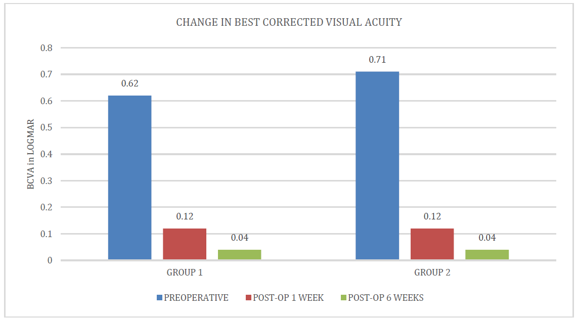
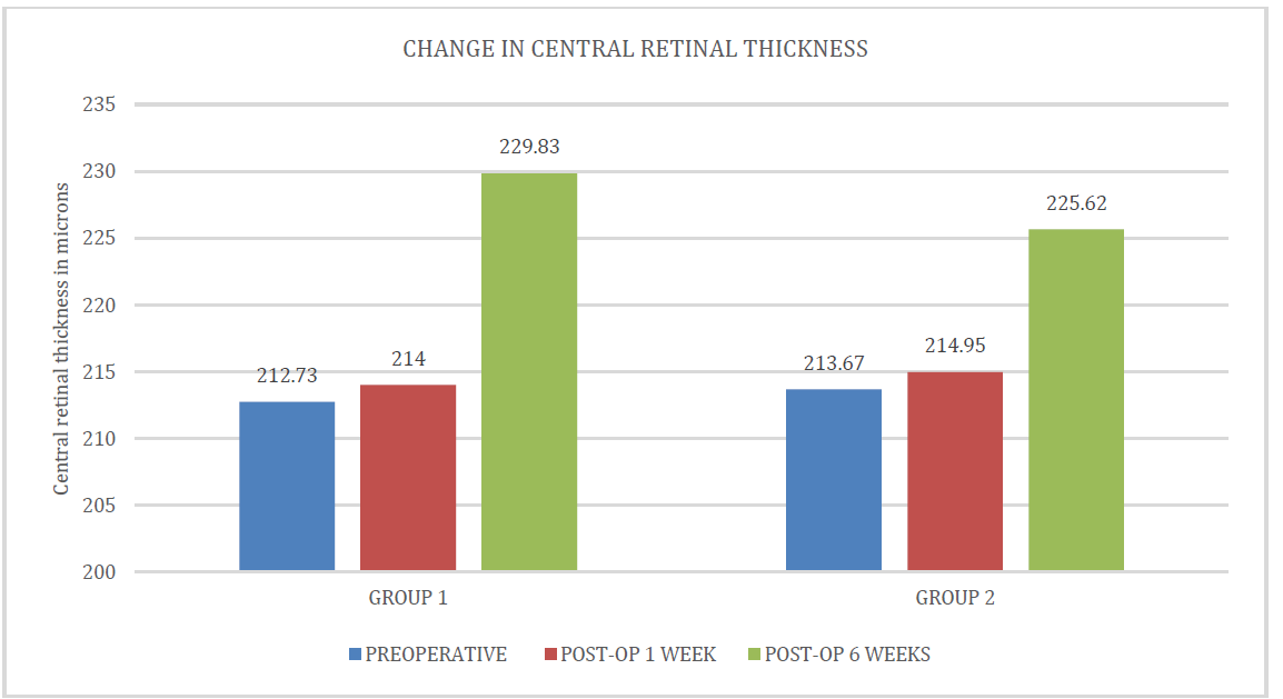
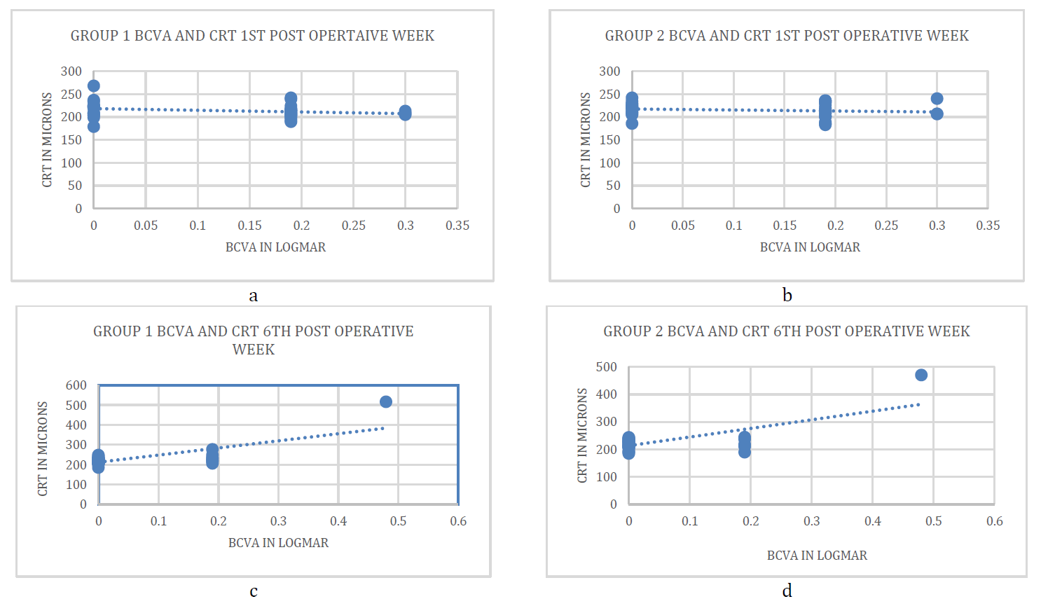
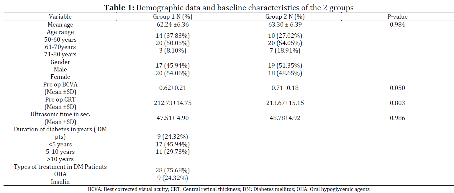
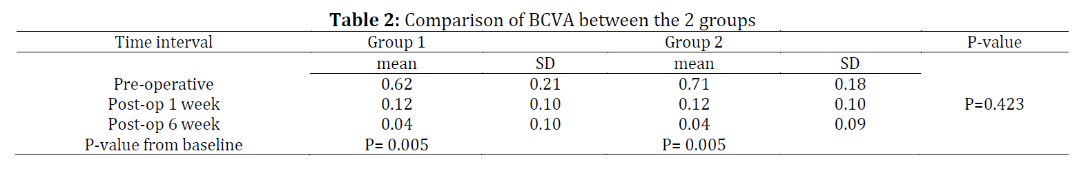
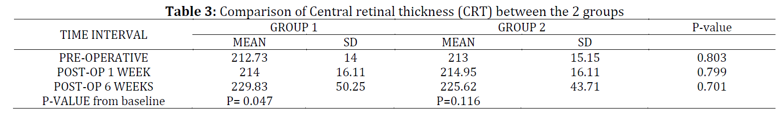
This study aims to determine the incidence of CME after uneventful phacoemulsification in diabetic patients without any signs of retinopathy and nondiabetic patients in early post-operative periods. The present study was a prospective, comparative, hospital-based study conducted from December 2019 to December 2020 in the Ophthalmology department of a tertiary care referral hospital in Eastern India. All patients aged between 50 to 80 years of either sex with cataract grade nuclear sclerosis II–III with or without cortical cataract and early posterior subcapsular cataract undergoing uncomplicated phacoemulsification surgery by a single surgeon were included in the study. The patients were divided into 2 groups. Group 1 consisted of Type 2 diabetes mellitus patients with cataracts with no retinopathy changes on funduscopy and group 2 included healthy non-diabetic patients with cataracts. Comprehensive baseline evaluation including central retinal thickness (CRT) measurement using optical coherence tomography was done in all cases. Best corrected visual acuity (BCVA) and CRT were assessed 1 week and 6 weeks following surgery. Seventy-four patients were included in the study. Both the groups consisted of 37 cases each. The mean age in group 1 and group 2 was 62.24 ± 6.36 and 63.30 ± 6.39 years respectively. One patient in each group developed clinical CME in the postoperative period. The incidence of CME in our study was 2.7%. Improvement in BCVA was comparable in both groups and statistically insignificant. An increase in CRT was found to be statistically significant (P= 0.047) in group 1 at the 6th week follow-up. Visual outcomes in diabetic patients without retinopathy are similar to normal patients following uncomplicated phacoemulsification surgery.
© 2022 The Authors. Published by IASE.
This is an
Keywords: Uneventful, Phacoemulsification, Cystoid macular edema, Diabetes, Retinopathy
Article History: Received 2 March 2022, Received in revised form 18 May 2022, Accepted 24 June 2022
Acknowledgment
No Acknowledgment.
Compliance with ethical standards
Conflict of interest: The author(s) declared no potential conflicts of interest with respect to the research, authorship, and/or publication of this article.
Citation:
Mehra L, Minj A, and Samant S et al. (2022). Occurrence of cystoid macular edema after uneventful phacoemulsification in diabetic patients without retinopathy: A hospital-based comparative study. International Journal of Advanced and Applied Sciences, 9(10): 59-65
Figures
Tables
----------------------------------------------
References (23)
- American Diabetes Association (2014). Diagnosis and classification of diabetes mellitus. Diabetes Care, 37(Supplement_1): S81-S90. https://doi.org/10.2337/dc14-S081 [Google Scholar] PMid:24357215
- Chu CJ, Johnston RL, Buscombe C, Sallam AB, Mohamed Q, and Yang YC (2016). United Kingdom pseudophakic macular edema study group risk factors and incidence of macular edema after cataract surgery: A database study of 81984 eyes. Ophthalmology, 123(2): 316-323. https://doi.org/10.1016/j.ophtha.2015.10.001 [Google Scholar] PMid:26681390
- Deshpande AC, Prabhudesai N, and Deshpande CV (2018). Incidence of clinical and subclinical cystoid macular edema in diabetic and non-diabetic patients after cataract surgery by means of optical coherence tomography. The Official Scientific Journal of Delhi Ophthalmological Society, 29(2): 39-43. https://doi.org/10.7869/djo.395 [Google Scholar]
- Dysli M, Rückert R, and Munk MR (2019). Differentiation of underlying pathologies of macular edema using spectral domain optical coherence tomography (SD-OCT). Ocular Immunology and Inflammation, 27(3): 474-483. https://doi.org/10.1080/09273948.2019.1603313 [Google Scholar] PMid:31184556
- Georgopoulos GT, Papaconstantinou D, Niskopoulou M, Moschos M, Georgalas I, and Koutsandrea C (2008). Foveal thickness after phacoemulsification as measured by optical coherence tomography. Clinical Ophthalmology, 2(4): 817-820. https://doi.org/10.2147/OPTH.S4031 [Google Scholar] PMid:19668435 PMCid:PMC2699786
- Guliani BP, Agarwal I, and Naik MP (2019). Effect of uncomplicated cataract surgery on central macular thickness in diabetic and non-diabetic subjects. Journal of Ophthalmic and Vision Research, 14(4): 442-447. https://doi.org/10.18502/jovr.v14i4.5447 [Google Scholar]
- Hartnett ME, Tinkham N, Paynter L, Geisen P, Rosenberg P, Koch G, and Cohen KL (2009). Aqueous vascular endothelial growth factor as a predictor of macular thickening following cataract surgery in patients with diabetes mellitus. American Journal of Ophthalmology, 148(6): 895-901. https://doi.org/10.1016/j.ajo.2009.07.014 [Google Scholar] PMid:19837384 PMCid:PMC2787768
- Hayashi K, Igarashi C, Hirata A, and Hayashi H (2009). Changes in diabetic macular oedema after phacoemulsification surgery. Eye, 23(2): 389-396. https://doi.org/10.1038/sj.eye.6703022 [Google Scholar] PMid:17962820
- Hee MR, Puliafito CA, Duker JS, Reichel E, Coker JG, Wilkins JR, and Fujimoto JG (1998). Topography of diabetic macular edema with optical coherence tomography. Ophthalmology, 105(2): 360-370. https://doi.org/10.1016/S0161-6420(98)93601-6 [Google Scholar]
- Katsimpris JM, Petropoulos IK, Zoukas G, Patokos T, Brinkmann CK, and Theoulakis PE (2012). Central foveal thickness before and after cataract surgery in normal and in diabetic patients without retinopathy. Klinische Monatsblätter für Augenheilkunde, 229(04): 331-337. https://doi.org/10.1055/s-0031-1299215 [Google Scholar] PMid:22495998
- Linebarger EJ, Hardten DR, Shah GK, and Lindstrom RL (1999). Phacoemulsification and modern cataract surgery. Survey of Ophthalmology, 44(2): 123-147. https://doi.org/10.1016/S0039-6257(99)00085-5 [Google Scholar]
- Loewenstein A and Zur D (2010). Postsurgical cystoid macular edema. Developments in Ophthalmology, 47: 148-159. https://doi.org/10.1159/000320078 [Google Scholar] PMid:20703048
- Miyake K and Ibaraki N (2002). Prostaglandins and cystoid macular edema. Survey of Ophthalmology, 47: 203-218. https://doi.org/10.1016/S0039-6257(02)00294-1 [Google Scholar]
- Nicholas S, Riley A, Patel H, Neveldson B, Purdie G, and Wells AP (2006). Correlations between optical coherence tomography measurement of macular thickness and visual acuity after cataract extraction. Clinical and Experimental Ophthalmology, 34(2): 124-129. https://doi.org/10.1111/j.1442-9071.2006.01169.x [Google Scholar] PMid:16626425
- Oyewole K, Tsogkas F, Westcott M, and Patra S (2017). Benchmarking cataract surgery outcomes in an ethnically diverse and diabetic population: final post-operative visual acuity and rates of post-operative cystoid macular oedema. Eye, 31(12): 1672-1677. https://doi.org/10.1038/eye.2017.96 [Google Scholar] PMid:28643796 PMCid:PMC5733300
- Pukl SS, Valentinčič VN, Urbančič M, Grčar II, Grčar R, Pfeifer V, and Petrovič GM (2017). Visual acuity, retinal sensitivity, and macular thickness changes in diabetic patients without diabetic retinopathy after cataract surgery. Journal of Diabetes Research, 2017: 3459156. https://doi.org/10.1155/2017/3459156 [Google Scholar] PMid:28243608 PMCid:PMC5294376
- Puliafito CA, Hee MR, Lin CP, Reichel E, Schuman JS, Duker JS, and Fujimoto JG (1995). Imaging of macular diseases with optical coherence tomography. Ophthalmology, 102(2): 217-229. https://doi.org/10.1016/S0161-6420(95)31032-9 [Google Scholar]
- Singhi A and Baishya KB (2017). A study on changes in macular thickness after cataract surgery in diabetic patients using optical coherence tomography (OCT). International Journal of Science and Research, 6(9): 1432-1437. [Google Scholar]
- Ursell PG, Spalton DJ, Whitcup SM, and Nussenblatt RB (1999). Cystoid macular edema after phacoemulsification: Relationship to blood–aqueous barrier damage and visual acuity. Journal of Cataract and Refractive Surgery, 25(11): 1492-1497. https://doi.org/10.1016/S0886-3350(99)00196-0 [Google Scholar]
- Von Jagow B, Ohrloff C, and Kohnen T (2007). Macular thickness after uneventful cataract surgery determined by optical coherence tomography. Graefe's Archive for Clinical and Experimental Ophthalmology, 245(12): 1765-1771. https://doi.org/10.1007/s00417-007-0605-6 [Google Scholar] PMid:17619896
- Wang KY and Cheng CK (2014). Central retinal thickness changes and visual outcomes following uncomplicated small-incision phacoemulsification cataract surgery in diabetic without retinopathy patients and nondiabetic patients. Taiwan Journal of Ophthalmology, 4(1): 33-39. https://doi.org/10.1016/j.tjo.2014.01.001 [Google Scholar]
- Yonekawa Y and Kim IK (2012). Pseudophakic cystoid macular edema. Current Opinion in Ophthalmology, 23(1): 26-32. https://doi.org/10.1097/ICU.0b013e32834cd5f8 [Google Scholar] PMid:22134362
- Yoon DH, Kang DJ, Kim MJ, and Kim HK (2018). New observation of microcystic macular edema as a mild form of cystoid macular lesions after standard phacoemulsification: Prevalence and risk factors. Medicine, 97(15): e0355. https://doi.org/10.1097/MD.0000000000010355 [Google Scholar] PMid:29642179 PMCid:PMC5908588