
International
ADVANCED AND APPLIED SCIENCES
EISSN: 2313-3724, Print ISSN:2313-626X
Frequency: 12
![]()
Volume 6, Issue 7 (July 2019), Pages: 89-98

----------------------------------------------
Original Research Paper
Title: Enhanced feature extraction technique for brain MRI classification based on Haar wavelet and statistical moments
Author(s): Zahid Ullah 1, Su-Hyun Lee 1, *, Muhammad Fayaz 2
Affiliation(s):
1Department of Computer Engineering, Changwon National University, Changwon, South Korea
2Department of Computer Engineering, Jeju National University, Jeju, South Korea

* Corresponding Author.
 Corresponding author's ORCID profile: https://orcid.org/0000-0001-6966-1569
Digital Object Identifier:
https://doi.org/10.21833/ijaas.2019.07.012
Abstract:
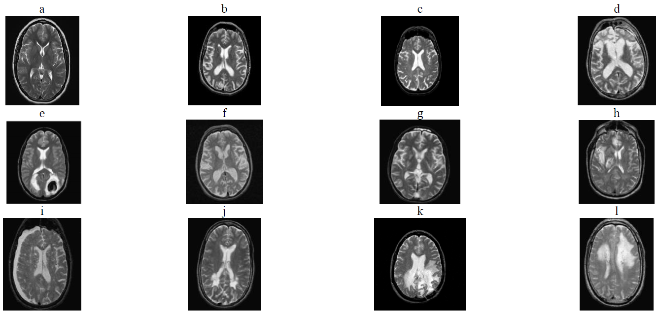
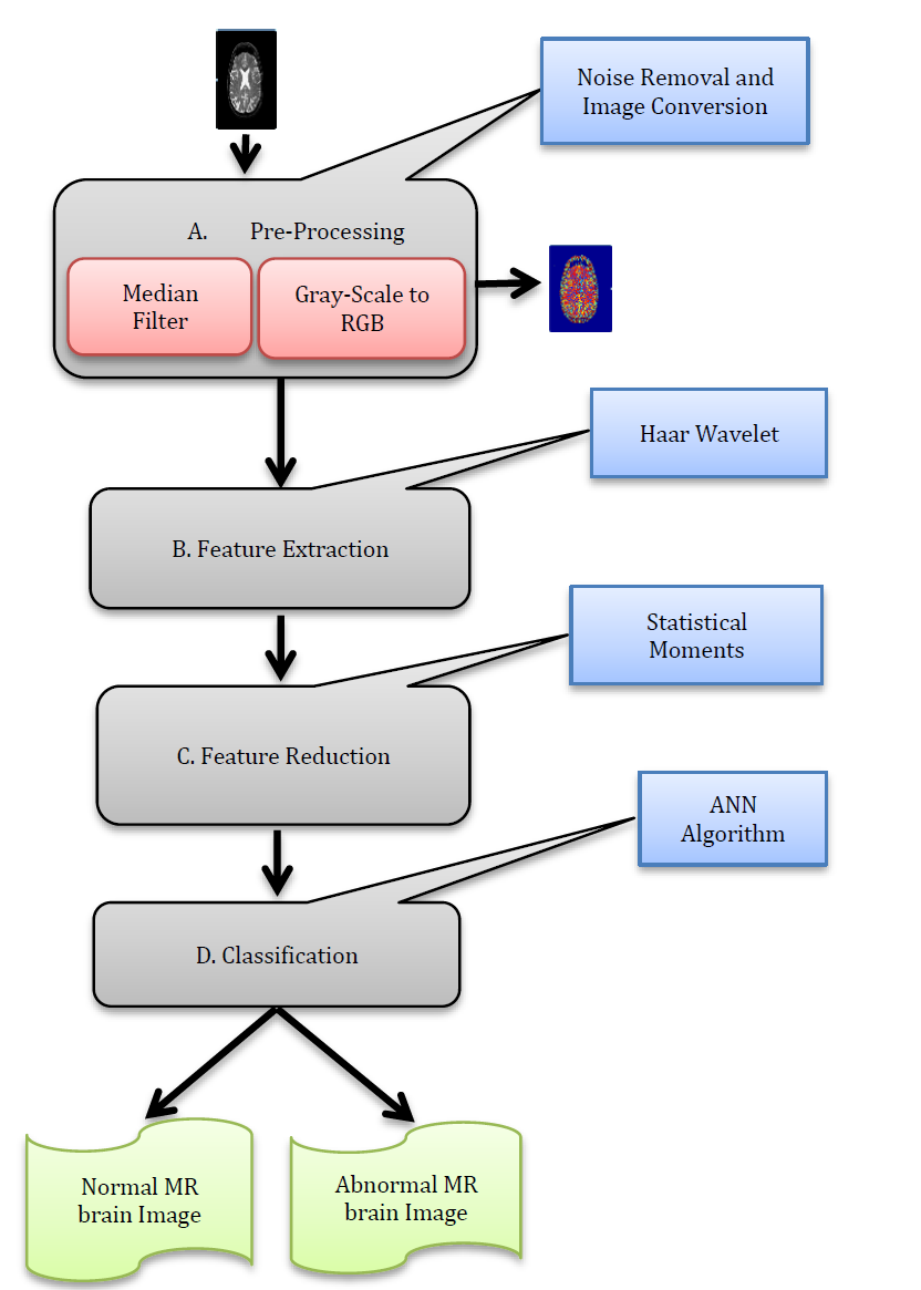
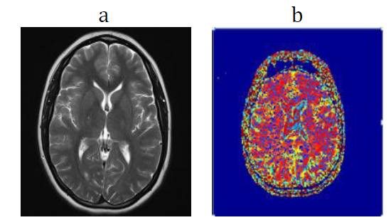
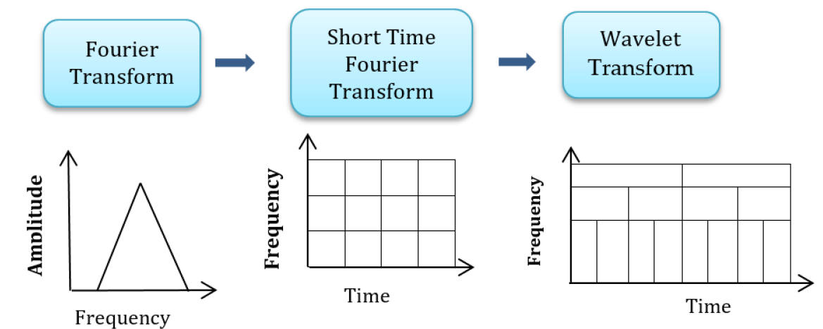
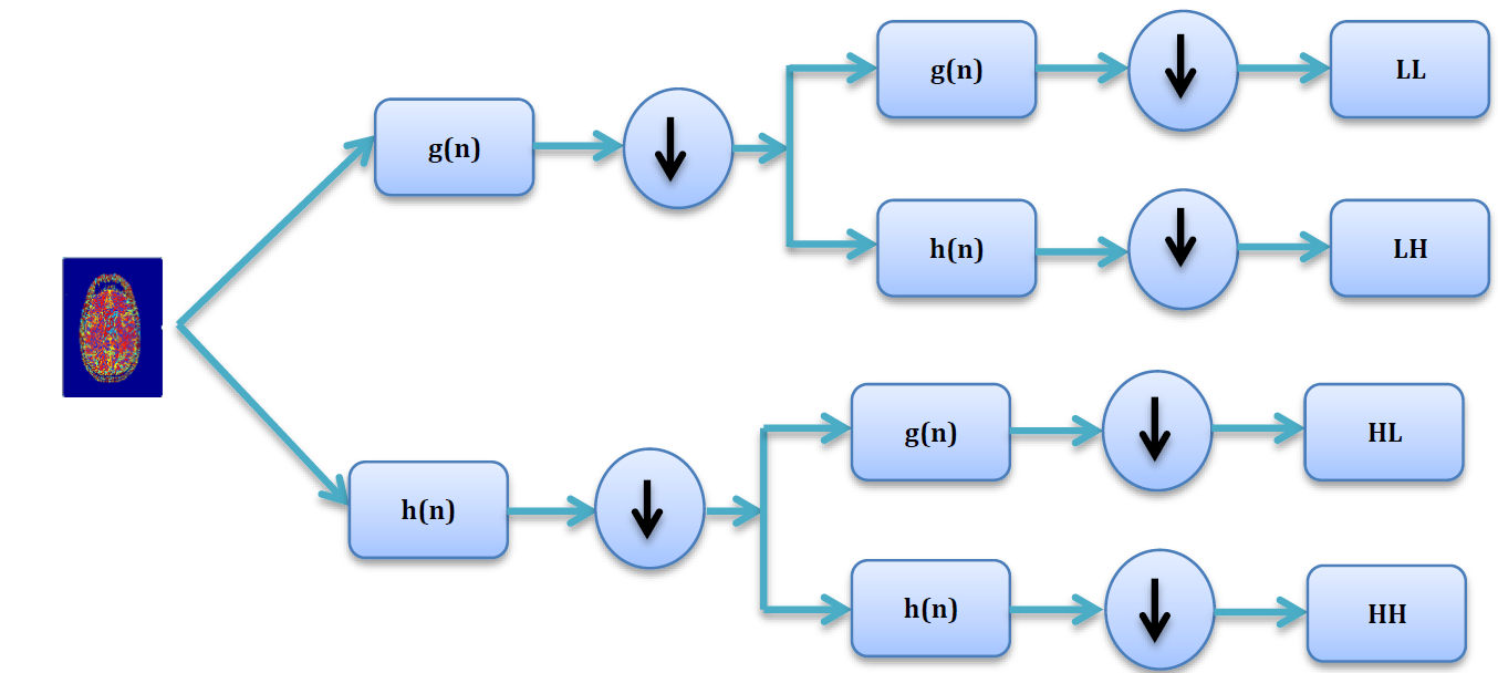
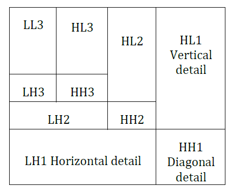
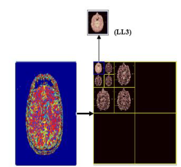
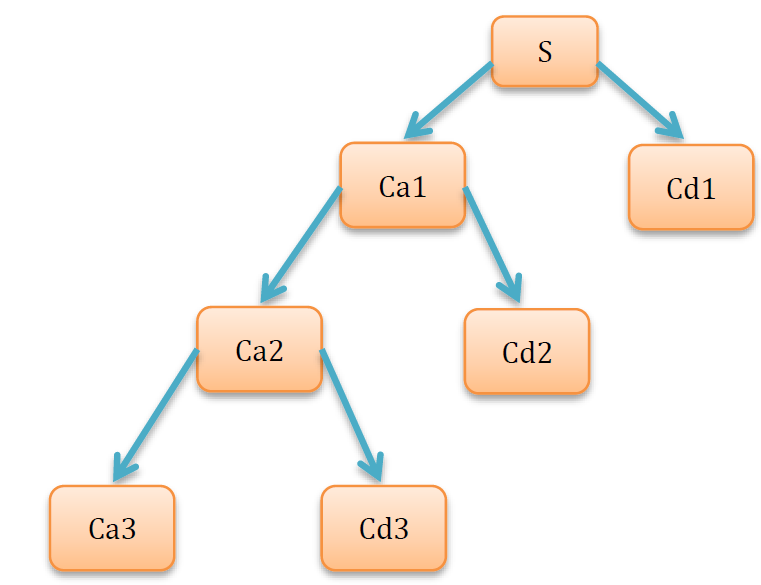
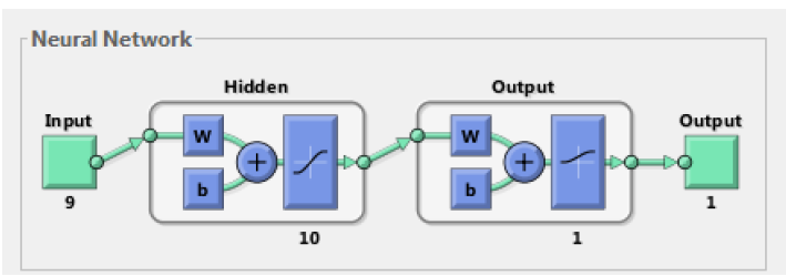
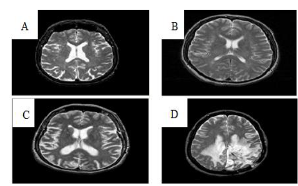
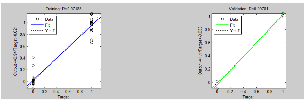
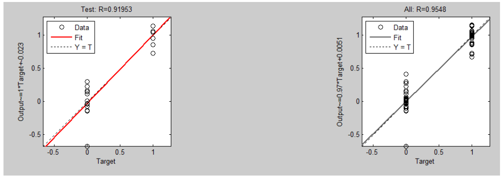
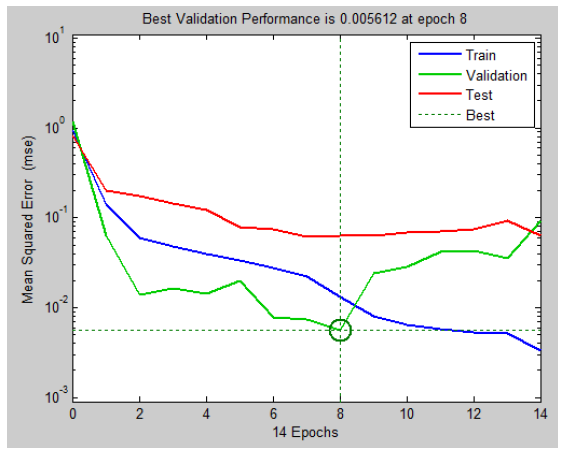
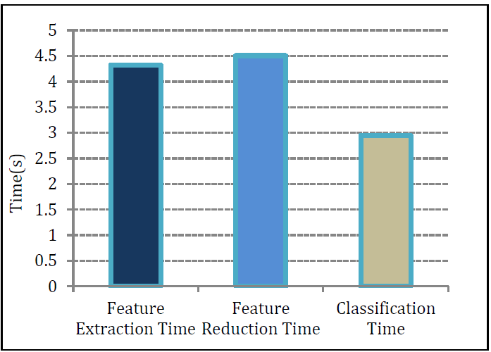
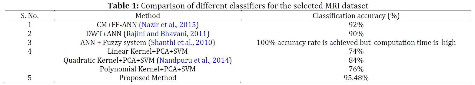
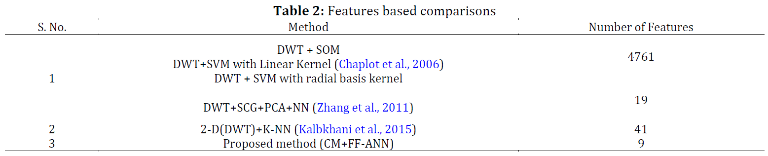
Many methods have been proposed to classify the MR brain images automatically. We have proposed a method based on a Neural Network (NN) to classify the normality and abnormality of a given MR brain image. This method first employs a median filter to minimize the noise from the image and converted the image to RGB. Then applies the technique of Discrete Wavelet Transform (DWT) to extract the important features from the image and color moments have been employed in the feature reduction stage to reduce the dimension of the features. The reduced features are sent to Feed-Forward Artificial neural network (FF-ANN) to discriminate the normal and abnormal MR brain images. We applied this proposed method on 70 images (45 normal, 25 abnormal). The accuracy of the proposed method of both training and testing images are 95.48%, while the computation time for feature extraction, feature reduction, and neural network classifier is 4.3216s, 4.5056s, and 1.4797s, respectively.
© 2019 The Authors. Published by IASE.
This is an
Keywords: MRI classification, Discrete wavelet transform, Color moments, Principal component analysis, Feature extraction, Approximation component, Artificial neural network
Article History: Received 14 January 2019, Received in revised form 20 May 2019, Accepted 25 May 2019
Acknowledgement:
No Acknowledgement.
Compliance with ethical standards
Conflict of interest: The authors declare that they have no conflict of interest.
Citation:
Ullah Z, Lee SH, and Fayaz M (2019). Enhanced feature extraction technique for brain MRI classification based on Haar wavelet and statistical moments. International Journal of Advanced and Applied Sciences, 6(7): 89-98
Figures
Fig. 1 Fig. 2 Fig. 3 Fig. 4 Fig. 5 Fig. 6 Fig. 7 Fig. 8 Fig. 9 Fig. 10 Fig. 11 Fig. 12 Fig. 13 Fig. 14
Tables
----------------------------------------------
References (34)
- Alfonse M and Salem ABM (2016). An automatic classification of brain tumours through MRI using support vector machine. Egyptian Computer Science Journal, 40(3): 11-21. [Google Scholar]
- Chaplot S, Patnaik LM, and Jagannathan NR (2006). Classification of magnetic resonance brain images using wavelets as input to support vector machine and neural network. Biomedical Signal Processing and Control, 1(1): 86-92. https://doi.org/10.1016/j.bspc.2006.05.002 [Google Scholar]
- Demirhan A and Güler İ (2011). Combining stationary wavelet transform and self-organizing maps for brain MR image segmentation. Engineering Applications of Artificial Intelligence, 24(2): 358-367. https://doi.org/10.1016/j.engappai.2010.09.008 [Google Scholar]
- El-Dahshan ESA, Hosny T, and Salem ABM (2010). Hybrid intelligent techniques for MRI brain images classification. Digital Signal Processing, 20(2): 433-441. https://doi.org/10.1016/j.dsp.2009.07.002 [Google Scholar]
- Fayaz M, Shah AS, Wahid F, and Shah A (2016). A robust technique of brain MRI classification using colour features and K-nearest neighbors algorithm. International Journal of Signal Processing, Image Processing and Pattern Recognition, 9(10): 11-20. https://doi.org/10.14257/ijsip.2016.9.10.02 [Google Scholar]
- Fletcher-Heath LM, Hall LO, Goldgof DB, and Murtagh FR (2001). Automatic segmentation of non-enhancing brain tumours in magnetic resonance images. Artificial Intelligence in Medicine, 21(1-3): 43-63. https://doi.org/10.1016/S0933-3657(00)00073-7 [Google Scholar]
- Gonzalez RC and Woods RE (2009). Digital image processing. Pearson Education, London, UK. https://doi.org/10.1117/1.3115362 [Google Scholar]
- Joseph RP, Singh CS, and Manikandan M (2014). Brain tumour MRI image segmentation and detection in image processing. International Journal of Research in Engineering and Technology, 3(1): 1-5. https://doi.org/10.15623/ijret.2014.0313001 [Google Scholar]
- Kalbkhani H, Salimi A, and Shayesteh MG (2015). Classification of brain MRI using multi-cluster feature selection and KNN classifier. In the 2015 23rd Iranian Conference on Electrical Engineering. IEEE, Tehran, Iran: 93-98. https://doi.org/10.1109/IranianCEE.2015.7146189 [Google Scholar]
- Keerthana TK and Xavier S (2018). An intelligent system for early assessment and classification of brain tumour. In the 2018 2nd International Conference on Inventive Communication and Computational Technologies, IEEE, Coimbatore, India: 1265-1268. https://doi.org/10.1109/ICICCT.2018.8473297 [Google Scholar]
- Korolev S, Safiullin A, Belyaev M, and Dodonova Y (2017). Residual and plain convolutional neural networks for 3D brain MRI classification. In the 2017 IEEE 14th International Symposium on Biomedical Imaging, IEEE, Melbourne, Australia: 835-838. https://doi.org/10.1109/ISBI.2017.7950647 [Google Scholar]
- Lavanyadevi R, Machakowsalya M, Nivethitha J, and Kumar AN (2017). Brain tumour classification and segmentation in MRI images using PNN. In the 2017 IEEE International Conference on Electrical, Instrumentation and Communication Engineering, IEEE, Karur, India: 1-6. https://doi.org/10.1109/ICEICE.2017.8191888 [Google Scholar]
- Logeswari T and Karnan M (2010). An improved implementation of brain tumour detection using segmentation based on soft computing. Journal of Cancer Research and Experimental Oncology, 2(1): 6-14. https://doi.org/10.1109/ICSAP.2010.55 [Google Scholar]
- Maitra M and Chatterjee A (2006). A Slantlet transform based intelligent system for magnetic resonance brain image classification. Biomedical Signal Processing and Control, 1(4): 299-306. https://doi.org/10.1016/j.bspc.2006.12.001 [Google Scholar]
- Maitra M and Chatterjee A (2008). Hybrid multiresolution Slantlet transform and fuzzy c-means clustering approach for normal-pathological brain MR image segregation. Medical Engineering and Physics, 30(5): 615-623. https://doi.org/10.1016/j.medengphy.2007.06.009 [Google Scholar] PMid:17698397
- Maniar S and Shah JS (2017). Classification of content based medical image retrieval using texture and shape feature with neural network. International Journal of Advances in Applied Sciences, 6(3): 368-374. [Google Scholar]
- Mathew AR and Anto PB (2017). Tumour detection and classification of MRI brain image using wavelet transform and SVM. In the 2017 International Conference on Signal Processing and Communication, IEEE, Coimbatore, India: 75-78. https://doi.org/10.1109/CSPC.2017.8305810 [Google Scholar] PMid:29173559
- Nandpuru HB, Salankar SS, and Bora VR (2014). MRI brain cancer classification using support vector machine. In the 2014 IEEE Students' Conference on Electrical, Electronics and Computer Science, IEEE, Bhopal, India: 1-6. https://doi.org/10.1109/SCEECS.2014.6804439 [Google Scholar]
- Nazir M, Wahid F, and Ali Khan S (2015). A simple and intelligent approach for brain MRI classification. Journal of Intelligent and Fuzzy Systems, 28: 1127-1135. https://doi.org/ 10.3233/IFS-141396 [Google Scholar]
- Rajini NH and Bhavani R (2011). Classification of MRI brain images using k-nearest neighbor and artificial neural network. In the 2011 International Conference on Recent Trends in Information Technology (ICRTIT), IEEE, Chennai, Tamil Nadu, India: 563-568. https://doi.org/10.1109/ICRTIT.2011.5972341 [Google Scholar]
- Saha C and Hossain MF (2017). MRI brain tumour images classification using K-means clustering, NSCT and SVM. In the 2017 4th IEEE Uttar Pradesh Section International Conference on Electrical, Computer and Electronics, IEEE, Mathura, India: 329-333. https://doi.org/10.1109/UPCON.2017.8251069 [Google Scholar]
- Saleh SR and Al-Bakry AM (2017). MRI images classification based on software agent. In the 2017 Annual Conference on New Trends in Information and Communications Technology Applications, IEEE, Baghdad, Iraq: 225-229. https://doi.org/10.1109/NTICT.2017.7976135 [Google Scholar]
- Shantanu G (2006). Inter-class relationships in text classification. Ph.D. Dissertation, Indian Institute of Technology, Bombay, India. [Google Scholar]
- Shanthi KJ, Sasikumar MN, and Kesavadas C (2010). Neuro-fuzzy approach toward segmentation of brain MRI based on intensity and spatial distribution. Journal of Medical Imaging and Radiation Sciences, 41(2): 66-71. https://doi.org/10.1016/j.jmir.2010.03.002 [Google Scholar] PMid:31051819
- Somasundaram K and Kalaiselvi T (2010). Fully automatic brain extraction algorithm for axial T2-weighted magnetic resonance images. Computers in Biology and Medicine, 40(10): 811-822. https://doi.org/10.1016/j.compbiomed.2010.08.004 [Google Scholar] PMid:20832783
- Suhaimi F and Htike ZZ (2018). Feature map size selection for fMRI classification on end-to-end deep convolutional neural network. International Journal of Advanced and Applied Sciences, 5(8): 95-103. https://doi.org/10.21833/ijaas.2018.08.012
- Ullah Z, Fayaz M, and Iqbal A (2016). Critical analysis of data mining techniques on medical data. International Journal of Modern Education and Computer Science, 8(2): 42-48. https://doi.org/10.5815/ijmecs.2016.02.05 [Google Scholar]
- Ullah Z, Lee SH, Khan MN, Fayaz M, and Iqbal MM (2018). Features reductions using colour moments and classification of brain MRI using K-NN. Technical Journal, 23(4). Available online at: https://bit.ly/30JVCKh
- Vasuda P and Satheesh S (2010). Improved fuzzy C-means algorithm for MR brain image segmentation. International Journal on Computer Science and Engineering, 2(5): 1713-1715. [Google Scholar]
- Wahid F, Ghazali R, Fayaz M, and Shah AS (2016). Using probabilistic classification technique and statistical features for brain magnetic resonance imaging (MRI) classification: An application of AI technique in bio-science. International Journal of Bio-Science and Bio-Technology, 8(6): 93-106. https://doi.org/10.14257/ijbsbt.2016.8.6.10 [Google Scholar]
- Westbrook C (2014). Handbook of MRI technique. John Wiley and Sons, Hoboken, USA. [Google Scholar]
- Zanaty EA and Aljahdali S (2011). Improving fuzzy algorithms for automatic image segmentation. In the 2011 International Conference on Multimedia Computing and Systems, IEEE, Ouarzazate, Morocco: 1-6. https://doi.org/10.1109/ICMCS.2011.5945581 [Google Scholar]
- Zarandi MF, Zarinbal M, and Izadi M (2011). Systematic image processing for diagnosing brain tumours: A Type-II fuzzy expert system approach. Applied Soft Computing, 11(1): 285-294. https://doi.org/10.1016/j.asoc.2009.11.019 [Google Scholar]
- Zhang Y, Dong Z, Wu L, and Wang S (2011). A hybrid method for MRI brain image classification. Expert Systems with Applications, 38(8): 10049-10053. https://doi.org/10.1016/j.eswa.2011.02.012 [Google Scholar]