
International
ADVANCED AND APPLIED SCIENCES
EISSN: 2313-3724, Print ISSN:2313-626X
Frequency: 12
![]()
Volume 6, Issue 7 (July 2019), Pages: 29-35

----------------------------------------------
Original Research Paper
Title: Clinical and cardiologic assessment of Anatolian shepherd dogs with asymptomatic degenerative mitral valve disease
Author(s): Kursad Turgut 1, *, Amir Naseri 2, Mehmet Ege Ince 1, Havva Süleymanoğlu 1, Merve Ertan 1, Vedat Sağmanlıgil 1, Ismail Sen 2, 3
Affiliation(s):
1Faculty of Veterinary Medicine, Near East University, Nicosia, Northern Cyprus
2Faculty of Veterinary Medicine, Selcuk University, Konya, Turkey
3Faculty of Veterinary Medicine, Kyrgyz Turkish Manas University, Bishkek, Kyrgyzstan

* Corresponding Author.
 Corresponding author's ORCID profile: https://orcid.org/0000-0001-8725-8044
Digital Object Identifier:
https://doi.org/10.21833/ijaas.2019.07.004
Abstract:
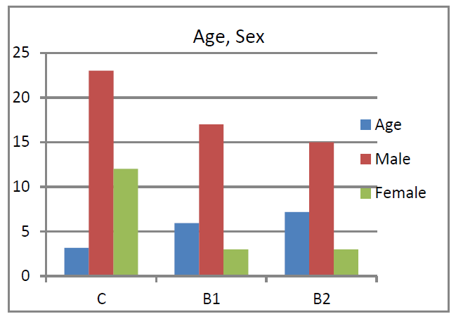
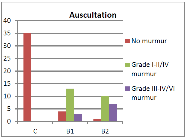
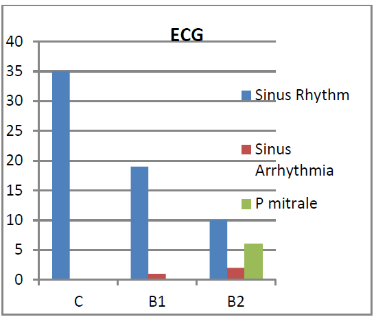
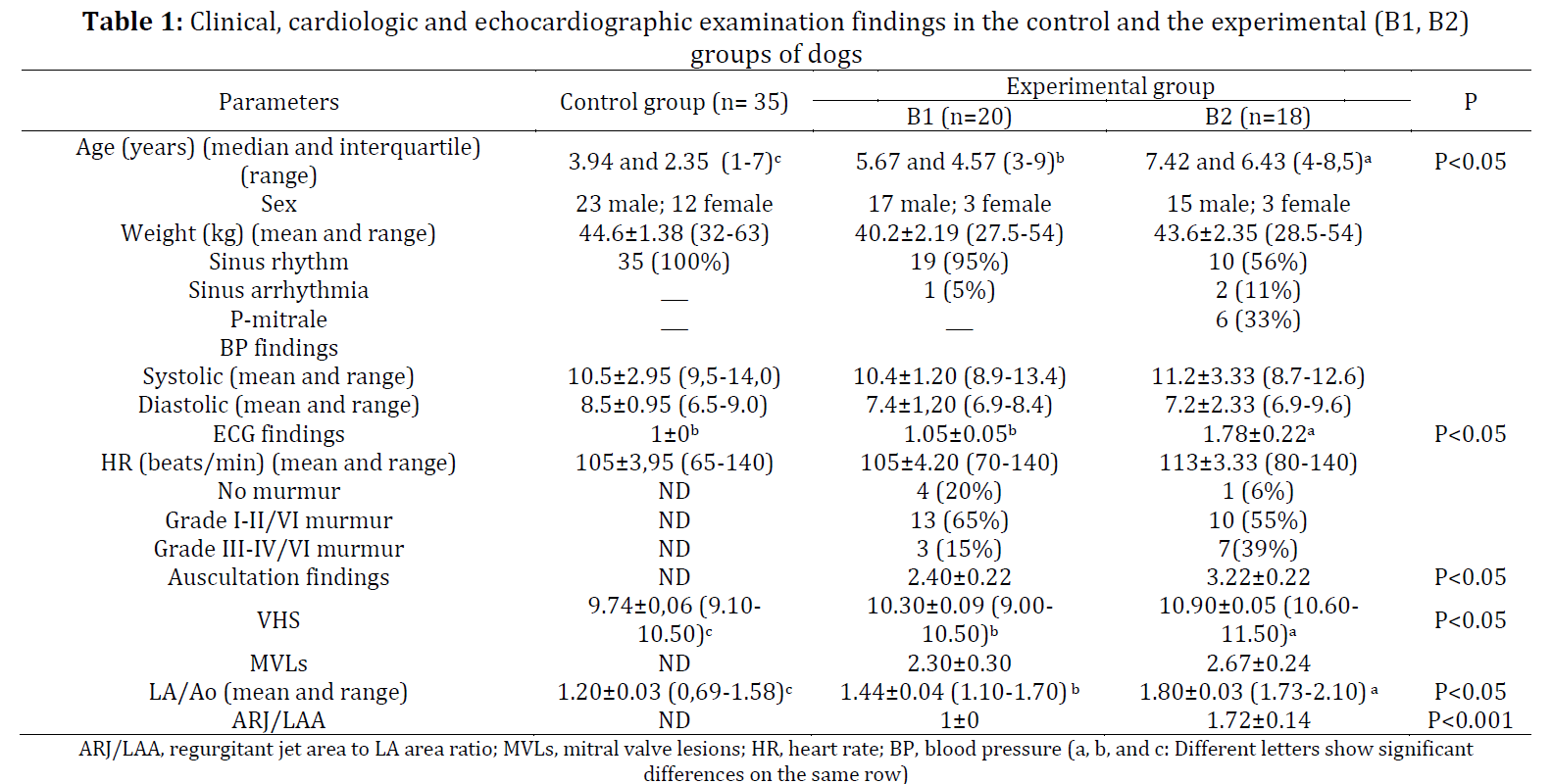
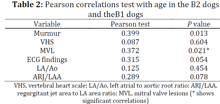
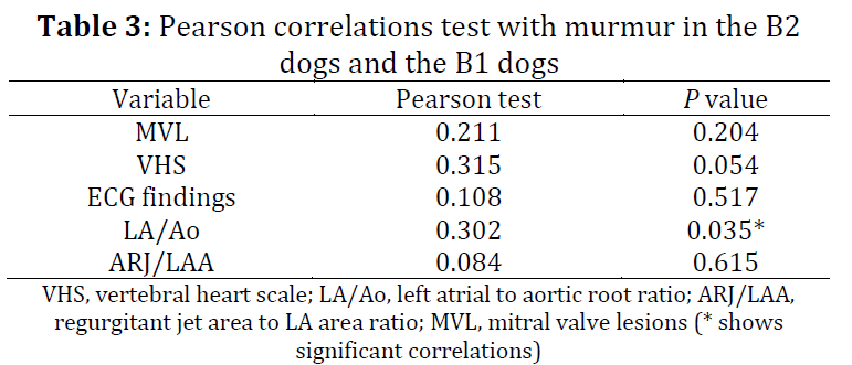
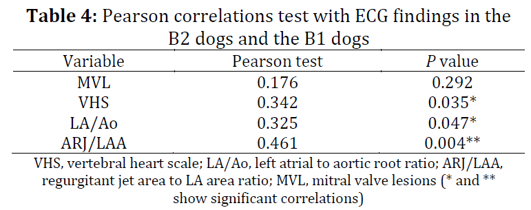
Degenerative mitral valve disease (DMVD) is the leading cause of cardiac disease and heart failure in the dog. Advanced age, breed and male gender are well-known risk factors for DMVD. The incidence of the disease in German Shepherds seems to be noteworthy. Early diagnosis of DMVD is related to the identification of a left apical systolic murmur, characteristic of MR in a dog. Dogs with DMVD had a low frequency of arrhythmias compared to other cardiac conditions. The goal of the study was (i) to evaluate the age and gender incidences of the asymptomatic Anatolian Shepherd Dogs (ASHs) with DMVD and, (ii) to investigate the importance of its clinical, radiological, electrocardiographic (ECG) findings and the correlations of those with some echo cardio logical measurements. 35 healthy ASHs (control group) and 38 ASHs with DMVD (experimental group) were used as the materials. The severity of cardiac disease was classified according to the American College of Veterinary Internal Medicine (ACVIM) consensus statement. Thirty two dogs (84.2%) were males and 6 dogs (15.8%) were females in the experimental group. The median age, the intensity of heart murmur and the severity of mitral regurgitation (MR) of the B2 dogs were bigger (p <0.05) than that of the B1 dogs. There was a positive correlation (P<0.05) between age and mitral valve lesions (MVLs). The clinical examination assessed by cardiac auscultation (murmur) was not correlated to MVLs, VHS, ECG findings and ARJ/LAA (P>0.05). The intensity of murmur was correlated to left ventricle to aorta ratio (LA/Ao) and it was not correlated (P>0.05) to MVLs, vertebral heart scale (VHS), ECG findings and regurgitant jet area to LA area ratio (ARJ/LAA). The correlations between ECG findings and VHS, along with, LA/Ao and ARJ/LAA were positive (P <0.05). In conclusion, aging and male gender may have a significant impact on DMVD progression in ASHs. Assessment of higher murmur in group B2 might be related to the progressive severity of the illnesses. The prevalence of arrhythmia was low in asymptomatic ASHs with DMVD. P-mitrale was noteworthy.
© 2019 The Authors. Published by IASE.
This is an
Keywords: Anatolian shepherd dog, Clinical assessment, Degenerative mitral valve disease, Echocardiography
Article History: Received 14 February 2019, Received in revised form 28 April 2019, Accepted 30 April 2019
Acknowledgement:
We are thankful to Dr. Enver Yazar for excellent assistance to statistical analyses.
Compliance with ethical standards
Conflict of interest: The authors declare that they have no conflict of interest.
Ethical approval:
This study was approved by Ethic committee of Faculty of Veterinary Medicine, University of Selcuk (Permit number: 2012/053).
All applicable international, national, and/or institutional guidelines for the care and use of animals were followed.
Citation:
Turgut K, Naseri A, and Ince ME et al. (2019). Clinical and cardiologic assessment of Anatolian shepherd dogs with asymptomatic degenerative mitral valve disease. International Journal of Advanced and Applied Sciences, 6(7): 29-35
Figures
Tables
Table 1 Table 2 Table 3 Table 4
----------------------------------------------
References (36)
- Bernay F, Bland JM, Häggström J, Baduel L, Combes B, Lopez A, and Kaltsatos V (2010). Efficacy of spironolactone on survival in dogs with naturally occurring mitral regurgitation caused by myxomatous mitral valve disease. Journal of Veterinary Internal Medicine, 24(2): 331-341. https://doi.org/10.1111/j.1939-1676.2009.0467.x [Google Scholar] PMid:20102506
- Bonagura JD and Schober KE (2009). Can ventricular function be assessed by echocardiography in chronic canine mitral valve disease?. Journal of Small Animal Practice, 50(s1): 12-24. https://doi.org/10.1111/j.1748-5827.2009.00803.x [Google Scholar] PMid:19765216
- Borgarelli M and Buchanan JW (2012). Historical review, epidemiology and natural history of degenerative mitral valve disease. Journal of Veterinary Cardiology, 14(1): 93-101. https://doi.org/10.1016/j.jvc.2012.01.011 [Google Scholar] PMid:22386588
- Borgarelli M, Zini E, D'Agnolo G, Tarducci A, Santilli RA, Chiavegato D, and Häggström J (2004). Comparison of primary mitral valve disease in German Shepherd dogs and in small breeds. Journal of Veterinary Cardiology, 6(2): 27-34. https://doi.org/10.1016/S1760-2734(06)70055-8 [Google Scholar]
- Buchanan JW (1977). Chronic valvular disease (endocardiosis) in dogs. Advances in Veterinary Sciences and Comparative Medicine, 7: 75-106. [Google Scholar]
- Buchanan JW and Bücheler J (1995). Vertebral scale system to measure canine heart size in radiographs. Journal of The American Veterinary Medical Association, 206: 194-194. [Google Scholar]
- Chetboul V and Tissier R (2012). Echocardiographic assessment of canine degenerative mitral valve disease. Journal of Veterinary Cardiology, 14(1): 127-148. https://doi.org/10.1016/j.jvc.2011.11.005 [Google Scholar] PMid:22366573
- Chirife R, Feitosa GS, and Frankl WS (1975). Electrocardiographic detection of left atrial enlargement: Correlation of P wave with left atrial dimension by echocardiography. Heart, 37(12): 1281-1285. https://doi.org/10.1136/hrt.37.12.1281 [Google Scholar]
- Connell PS, Han RI, and Grande-Allen KJ (2012). Differentiating the aging of the mitral valve from human and canine myxomatous degeneration. Journal of Veterinary Cardiology, 14(1): 31-45. https://doi.org/10.1016/j.jvc.2011.11.003 [Google Scholar] PMid:22364720 PMCid:PMC3307912
- de Madron E (1992). Primary acquired mitral insufficiency in adult large breed dogs. In the 10th American College Veterinary İnternal Medicine Forum (ACVIM Forum), San Diego, USA: 608-609. [Google Scholar]
- de Madron E (1998). Unusual aspects of mitral valve disease in the dog. In the 16th American College Veterinary İnternal Medicine Forum (ACVIM Forum), San Diego, USA: 116-118. [Google Scholar]
- Garncarz M, Parzeniecka-Jaworska M, Jank M, and Łój M (2013). A retrospective study of clinical signs and epidemiology of chronic valve disease in a group of 207 Dachshunds in Poland. Acta Veterinaria Scandinavica, 55(1): 52-58. https://doi.org/10.1186/1751-0147-55-52 [Google Scholar] PMid:23844824 PMCid:PMC3723884
- Gordon SG, Saunders AB, and Wesselowski SR (2017). Asymptomatic canine degenerative valve disease: Current and future therapies. Veterinary Clinics: Small Animal Practice, 47(5): 955-975. https://doi.org/10.1016/j.cvsm.2017.04.003 [Google Scholar] PMid:28669433
- Häggström J, Höglund K, and Borgarelli M (2009). An update on treatment and prognostic indicators in canine myxomatous mitral valve disease. Journal of Small Animal Practice, 50(s1): 25-33. https://doi.org/10.1111/j.1748-5827.2009.00800.x [Google Scholar] PMid:19765217
- Hyun CB (2005). Mitral valve prolapse in cavalier King Charles spaniel: A review and case study. Journal of Veterinary Science, 6(1): 67-73. https://doi.org/10.4142/jvs.2005.6.1.67 [Google Scholar] PMid:15785126
- Jones TC and Zook BC (1965). Aging changes in the vascular system of animals. Annals of the New York Academy of Sciences, 127(1): 671-684. https://doi.org/10.1111/j.1749-6632.1965.tb49434.x [Google Scholar]
- Kogure K (1980). Pathology of chronic mitral valvular disease in the dog. Japanese Journal of Veterinary Science, 42(3): 323-335. https://doi.org/10.1292/jvms1939.42.323 [Google Scholar] PMid:7218618
- Ljungvall I, Rishniw M, Porciello F, Ferasi L, and Ohad DG (2014). Murmur intensity in small‐breed dogs with myxomatous mitral valve disease reflects disease severity. Journal of Small Animal Practice, 55(11): 545-550. https://doi.org/10.1111/jsap.12265 [Google Scholar] PMid:25213440
- Lopez-Alvarez J, Boswood A, Moonarmart W, Hezzell MJ, Lotter N, and Elliott J (2014). Longitudinal electrocardiographic evaluation of dogs with degenerative mitral valve disease. Journal of Veterinary Internal Medicine, 28(2): 393-400. https://doi.org/10.1111/jvim.12311 [Google Scholar] PMid:24494591 PMCid:PMC4857969
- Martin M (2007). Small animal ECGs: An introductory guide. 2nd Edition, Blackwell Publishing Ltd, Hoboken, USA. [Google Scholar]
- Matos JM and Glaus TM (2010). Medical treatment of canine heart failure. European Journal of Companion Animal Practice, 20(2): 171-176. [Google Scholar]
- McGinley JC, Berretta RM, Chaudhary K, Rossman E, Bratinov GD, Gaughan JP, and Margulies KB (2007). Impaired contractile reserve in severe mitral valve regurgitation with a preserved ejection fraction. European Journal of Heart Failure, 9(9): 857-864. https://doi.org/10.1016/j.ejheart.2007.05.013 [Google Scholar] PMid:17594913
- Pedersen D, Lorentzen KA, and Kristensen BØ (1999a). Echocardiographic mitral valve prolapse in cavalier King Charles spaniels: Epidemiology and prognostic significance for regurgitation. Veterinary Record, 144(12): 315-320. https://doi.org/10.1136/vr.144.12.315 [Google Scholar] PMid:10212505
- Pedersen HD and Häggström J (2000). Mitral valve prolapse in the dog: A model of mitral valve prolapse in man. Cardiovascular Research, 47(2): 234-243. https://doi.org/10.1016/S0008-6363(00)00113-9 [Google Scholar]
- Pedersen HD, Häggström J, Falk T, Mow T, Olsen LH, Iversen L, and Jensen AL (1999b). Auscultation in mild mitral regurgitation in dogs: observer variation, effects of physical maneuvers, and agreement with color doppler echocardiography and phonocardiography. Journal of Veterinary Internal Medicine, 13(1): 56-64. https://doi.org/10.1111/j.1939-1676.1999.tb02166.x [Google Scholar] PMid:10052065
- Petric AD (2015). Myxomatous mitral valve disease in dogs-An update and perspectives. Macedonian Veterinary Review, 38(1): 13-20. https://doi.org/10.14432/j.macvetrev.2014.11.026 [Google Scholar]
- Rasmussen CE, Falk T, Zois NE, Moesgaard SG, Häggström J, Pedersen HD, and Olsen LH (2012). Heart rate, heart rate variability, and arrhythmias in dogs with myxomatous mitral valve disease. Journal of Veterinary Internal Medicine, 26(1): 76-84. https://doi.org/10.1111/j.1939-1676.2011.00842.x [Google Scholar] PMid:22151356
- Rasmussen CE, Vesterholm S, Ludvigsen TP, Häggström J, Pedersen HD, Moesgaard SG, and Olsen LH (2011). Holter monitoring in clinically healthy Cavalier King Charles Spaniels, Wire‐haired Dachshunds, and Cairn Terriers. Journal of Veterinary Internal Medicine, 25(3): 460-468. https://doi.org/10.1111/j.1939-1676.2011.0707.x [Google Scholar] PMid:21418322
- Rishniw M and Erb HN (2000). Evaluation of four 2‐dimensional echocardiographic methods of assessing left atrial size in dogs. Journal of Veterinary Internal Medicine, 14(4): 429-435. https://doi.org/10.1111/j.1939-1676.2000.tb02252.x [Google Scholar] PMid:10935894
- Serfass P, Chetboul V, Sampedrano CC, Nicolle A, Benalloul T, Laforge H, and Tissier R (2006). Retrospective study of 942 small-sized dogs: Prevalence of left apical systolic heart murmur and left-sided heart failure, critical effects of breed and sex. Journal of Veterinary Cardiology, 8(1): 11-18. https://doi.org/10.1016/j.jvc.2005.10.001 [Google Scholar] PMid:19083332
- Terzo E, Di Marcello M, Mcallıster H, Glazier B, Lo Coco D, Locatelli C, and Brambilla PG (2009). Echocardiographic assessment of 537 dogs with mitral valve prolapse and leaflet involvement. Veterinary Radiology and Ultrasound, 50(4): 416-422. https://doi.org/10.1111/j.1740-8261.2009.01559.x [Google Scholar]
- Thrusfield MV, Aitken CGG, and Darker PGG (1985). Observations on breed and sex in relation to canine heart valve incompetence. Journal of Small Animal Practice, 26(12): 709-717. https://doi.org/10.1111/j.1748-5827.1985.tb02199.x [Google Scholar]
- Trafny DJ, Freeman LM, Bulmer BJ, MacGregor JM, Rush JE, Meurs KM, and Oyama MA (2012). Auscultatory, echocardiographic, biochemical, nutritional, and environmental characteristics of mitral valve disease in Norfolk terriers. Journal of Veterinary Cardiology, 14(1): 261-267. https://doi.org/10.1016/j.jvc.2011.10.002 [Google Scholar] PMid:22364691
- Turgut K (2017). Klinik kedi ve köpek kardiyolojisi. Nobel Tıp Kitabevleri, Istanbul, Turkey. [Google Scholar]
- Ware W (2011). Cardiovascular disease in small animal medicine. CRC Press, Boca Raton, USA. https://doi.org/10.1201/b15177 [Google Scholar]
- Whitney JG (1974). Observations on the effect of age on the severity of heart valve lesions in the dog. Journal of Small Animal Practice, 15(8): 511-522. https://doi.org/10.1111/j.1748-5827.1974.tb06529.x [Google Scholar] PMid:4469562